Herpes zoster: Understanding the disease, its treatment, and prevention

Herpes zoster: Understanding the disease, its treatment, and prevention

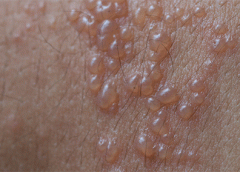
Herpes zoster (HZ, also called shingles) is a painful condition that produces a maculopapular and vesicular rash. Usually, the rash appears along a single dermatome (band) around one side of the body or face.

In most cases, pain, tingling, burning, or itching occurs a few days before the rash. Next, blisters form, scabbing over in 7 to 10 days. In rare cases, the rash is widespread, resembling varicella zoster (VZ, or chickenpox) rash. Pain can range from mild to severe and may be dull, burning, or gnawing. It may last weeks, months, or even years after the blisters heal. Shingles on the face may impair vision or hearing. (more…)

Read More

2016 Journal: November – December Vol. 5 No. 6

Wound Care Advisor Journal 2016 Nov/Dec Vol. 5 No. 6

Herpes zoster: Understanding the disease, its treatment, and prevention


Herpes zoster (HZ, also called shingles) is a painful condition that produces a maculopapular and vesicular rash. Usually, the rash appears along a single dermatome (band) around one side of the body or face. In most cases, pain, tingling, burning, or itching occurs a few days before the rash. Next, blisters form, scabbing over in 7 to 10 days. In…
read more

Frequently asked questions about support surfaces

The National Pressure Ulcer Advisory Panel (NPUAP) describes support surfaces as “specialized devices for pressure redistribution designed for management of tissue loads, microclimate, and/or other therapeutic functions.” These devices include specialized mattresses, mattress overlays, chair cushions, and pads used on transport stretchers, operating room (OR) tables, examination or procedure tables, and gurneys. Some support surfaces are part of an integrated…

Herpes zoster: Understanding the disease, its treatment, and prevention

Herpes zoster: Understanding the disease, its treatment, and prevention

Herpes zoster (HZ, also called shingles) is a painful condition that produces a maculopapular and vesicular rash. Usually, the rash appears along a single dermatome (band) around one side of the body or face. In most cases, pain, tingling, burning, or itching occurs a few days before the rash. Next, blisters form, scabbing over in 7 to 10 days. In…

Knowing when to ask for help

As a wound care expert, you’re probably consulted for every eruption, scrape, and opening in a patient’s skin. Occasionally during a patient assessment, you may scratch your head and ask yourself, “What is this? I’ve never seen anything like it.” Most wound care experts want to help heal everyone, and most of us love a challenge. But when should we…

Managing chronic venous leg ulcers — what’s the latest evidence?

Managing chronic venous leg ulcers — what’s the latest evidence?

Chronic venous leg ulcers (CVLUs) affect nearly 2.2 million Americans annually, including an estimated 3.6% of people over the age of 65. Given that CVLU risk increases with age, the global incidence is predicted to escalate dramatically because of the growing population of older adults. Annual CVLU treatment-related costs to the U.S. healthcare system alone are upwards of $3.5 billion,…

Ostomy documentation tips

General characteristics Document if the diversion is an intestinal or urinary ostomy, whether it’s temporary or permanent, and the location— abdominal quadrant, skin fold, umbilicus. (See Descriptor reference.)

Skin substitutes: Understanding product differences

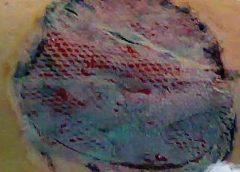
Skin substitutes (also called tissuebased products and dermal replacements) are a boon to chronic wound management when traditional therapies have failed. When selecting skin substitutes for their formularies, wound care professionals have many product options—and many decisions to make. Repair of skin defects has been a pressing concern for centuries. As early as the 15th century BC, Egyptian physicians chronicled…

2016 Journal: November – December Vol. 5 No. 6

Click here to access the digital edition

Read More

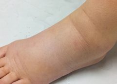
Lymphedema and lipedema: What every wound care clinician should know

Imagine you have a health condition that affects your life every day. Then imagine being told nothing can be done about it; you’ll just have to live with it. Or worse yet, your physician tells you the problem is “you’re just fat.”

Many people with lymphedema or lipedema have no idea their condition has a name or that many other people suffer from the same thing. Although lymphedema and lipedema can’t be cured, proper management and resources can help patients cope. This article improves your grasp of these conditions, describes how to recognize and manage them, and explains how to support your patients.

To understand lymphedema and lipedema, first you need to understand how the lymphatic system functions. It makes lymph, then moves it from tissues to the bloodstream. It also plays a major role in the immune system, aiding immune defense. In addition, it helps maintain normal fluid balance by promoting fluid movement from the interstitial tissues back to the venous circulation. (See Lymphatic system: Four major functions.)

If the lymphatic system is impaired from a primary (hereditary or congenital) condition or a secondary problem, lymphedema can result. In this chronic, potentially progressive, and incurable condition, protein-rich fluid accumulates in the interstitial tissues.

Lymphedema basics

Lymphedema occurs in four stages.

Stage 0. During this stage (also called the subclinical or latency stage), transport capacity of the lymphatic system decreases but remains sufficient to manage normal lymphatic loads. Signs and symptomsaren’t evident and can be measured only by sensitive instruments, such as bioimpedance spectroscopy and optoelectronic volumetry. Without such instruments to quantify volume changes, diagnosis may rest on subjective complaints.

In this stage, limited functional reserve of the lymphatic system leads to a fragile balance between subnormal transport capacity and lymphatic loads. Added stress on the lymphatic system (as from extended heat or cold exposure, injury, or infection) may cause progression to stage 1.

Providing appropriate patient information and education, especially after surgery, can dramatically reduce the risk that lymphedema will progress to a more serious stage.

Stage 1. Considered the spontaneously reversible stage, stage 1 is marked by softtissue pliability without fibrotic changes. Pitting can be induced easily. In early stage 1, limb swelling may recede over – night. With proper management, the patient can expect the extremity to decrease to a normal size compared to that of the uninvolved limb. Otherwise, lymphedema is likely to progress to stage 2.

Stage 1 lymphedema may be hard to distinguish from edemas from other causes. Clinicians must rely on the patient history and monitor for swelling resolution with conventional management, such as compression and elevation, or note if swelling persists despite these standard interventions.

Stage 2. Sometimes called the spontaneously irreversible stage, stage 2 is identified mainly from tissue proliferation and subsequent fibrosis (called lymphostatic fibrosis). The fluid component can be removed spontaneously, but removal of the increased tissue proliferation (initially irreversible) takes more time. Tissue proliferation stems from long-standing accumulation of protein-rich fluid; over time, the tissue hardens and pitting is hard to induce. In many cases, swelling volume increases, exacerbating the already compromised local immune defense.

Consequently, infections (particularly cellulitis) are common; these, in turn, increase the volume of the affected area. Proper treatment can reduce volume.

With proper care (complete decongestive therapy [CDT]), lymphedema can stabilize during stage 2. But patients with chronic or recurrent infections are likely to progress to stage 3.

Stage 3. Also called lymphostatic elephantiasis, this stage is marked by further fluid volume increases and progression of tissue changes. Lymphostatic fibrosis becomes firmer and other skin alterations may occur, including papillomas, cysts, fistulas, hyperkeratosis, fungal infections, and ulcers. Pitting may be present. Natural skinfolds deepen (especially those of the dorsum of the wrist or ankle) and, in many cases, cellulitis recurs.

If lymphedema management starts during this stage, reduction can still occur. Even in extreme cases, with proper care and patient adherence to treatment, lymphostatic elephantiasis can be reduced so the leg is a normal or near-normal size.

Assessment and diagnosis

A thorough physical examination is the gold standard for diagnosing lymphedema. A complete patient history, body-systems review, inspection, and palpation can help determine if edema is lymphedema.

Clinically, the only test with proven reliability and validity in diagnosing lymphedema is the Stemmer sign. Fibrotic changes associated with lymphedema can lead to thickened skin over the proximal phalanges of the toes or fingers. If you can’t tent or pinch the skin on the involved extremity, lymphedema is present (a positive Stemmer sign). However, a negative finding (soft, pliable tissue) doesn’t rule out  lymphedema because the condition may be in an early stage, before tissue proliferation and fibrosis have set in.

Management

Although incurable, lymphedema can be managed successfully through CDT. This approach involves proper identification of lymphedema, manual lymph drainage, skin and nail care, patient education, compression, and exercise.

CDT has two phases:

Phase I, the intensive phase, continues until the extremity has decongested or reached a plateau. The clinician provides treatments and educates the patient about all aspects of CDT to prepare him or her for phase II. Phase I can last several weeks to several months depending on lymphedema severity.

Phase II, the maintenance phase, begins once the extremity has decongested or plateaued. This phase still focuses on CDT, but now the patient, not the clinician, is responsible for all care. The goal is to reduce limb size while enabling the patient to become self-sufficient in managing lymphedema. Although CDT can bring significant improvements in limb size, skin quality, and function, patients must remember that phase II continues lifelong. Be sure to provide education about ongoing self-management strategies.

Lipedema: The disease they call “fat”

Lipedema is a painful disorder of fat deposition. Pathologic deposition of fatty tissue (usually below the waist) leads to progressive leg enlargement. Like lymphedema, lipedema is incurable but manageable. Unless managed properly, lipedema can reduce mobility, interfere with activities of daily living, and lead to secondary lymphedema. (See Lipedema stages.)

Lipedema commonly is misdiagnosed as lymphedema. However, lymphedema involves protein-rich fluid, whereas lip edema is a genetically mediated fat disorder. Because lipedema resists diet and exercise, it can lead to psychosocial complications. Lipedema occurs almost exclusively in women; typically, onset occurs between puberty and age 30. One unpublished epidemiologic study puts lip edema incidence in females at 11%. Some patients have a combination of lipedema and lymphedema. (See Viewing lipolymphedema.)

Assessment and diagnosis

As with lymphedema, lipedema diagnosis rests on clinical presentation. Lipedema characteristics include bilateral and symmetrical involvement, absence of pitting (because lipedema isn’t a fluid disorder), soft and pliable skin, and filling of the retromalleolar sulcus (called the fat pad sign.)

Key signs and symptoms include:

• feeling of heaviness in the legs (aching dysesthesia)

• easy bruising

• sensitivity to touch (called “painful fat syndrome”)

• orthostatic edema

• oatmeal-like changes to skin texture.

Nearly half of lipedema patients are overweight or obese, but many appear of normal weight from the waist up. Essentially, the upper and lower extremities don’t match. The lower extremities typically show fatty deposits extending from the iliac crest to the ankles, sparing the feet. (See Lipedema patterns.)

Management

Lipedema is best  managed through weight control, as additional weight gain through adipose tissue tends to deposit in the legs. For patients with concomitant lymphedema (lipolymphedema), modified CDT helps reduce and manage lymphatic compromise. To address excess fat deposition, newer “wet” liposuction techniques have proven beneficial. These techniques gently detach adipose cells from the tissue, helping to preserve connective tissue and lymphatic vessels.

Know what to look for

In both lymphedema and lipedema, early identification and proper diagnosis are key. (See Differentiating lymphedema and lipedema.) A thorough history and physical exam will likely lead to an accurate diagnosis, if clinicians know what to look for. Proper diagnosis and treatment can prevent expensive and ineffective interventions, which can negatively affect both the patient’s condition and psychological well being.

Heather Hettrick is an associate professor at Nova Southeastern University, Department of Physical Therapy in Fort Lauderdale, Florida.

Selected references

Fat Disorders Research Society. Lipedema description.

Fife CE, Maus EA, Carter MJ. Lipedema: a frequently misdiagnosed and misunderstood fatty deposition syndrome. Adv Skin Wound Care. 2010;23(2):81-92

Herbst KL. Rare adipose disorders (RADS) masquerading as obesity. Acta Pharmacol Sin. 2012;33(2):155-72.

Lipedema Project.

National Lymphedema Network. Position papers.

Schmeller W, Hueppe M, Meier-Vollrath I. Tumescent liposuction in lipoedema yields good long-term results. Br J Dermatol. 2012;166(1):161-8.

Zuther J. A closer look at lipedema and the effects on the lymphatic system. December 13, 2012. lymphedemablog.com/2012/12/13/a-closer-look-at-lipedema-and-the-effects-on-the-lymphatic-system/

Zuther J. Stages of lymphedema. October 3, 2012.

Read More

Understanding NPUAP’s updates to pressure ulcer terminology and staging

On April 13, 2016, the National Pressure Ulcer Advisory Panel (NPUAP) announced changes in pressure ulcer terminology and staging definitions. Providers can adapt NPUAP’s changes for their clinical practice and documentation, but it’s important to note that, as of press time, the Centers for Medicare & Medicaid Services (CMS) has not adopted the changes. This means that providers can’t use NPUAP’s updates when completing CMS assessment forms, such as the Minimum Data Set (MDS) or Outcome and Assessment Information Set (OASIS). Instead, they must code the CMS assessment forms according to current CMS instructions and definitions. In addition, there is no ICD-10 code for pressure injury. (more…)

Read More

How to manage peristomal skin problems

For an ostomy pouching system to adhere properly, the skin around the stoma must be dry and intact. Otherwise, peristomal skin problems and skin breakdown around the stoma may occur. In fact, these problems are the most common complications of surgical stomas. They can worsen the patient’s pain and discomfort, diminish quality of life, delay rehabilitation, increase use of ostomy supplies, and raise healthcare costs.

Peristomal skin problems also perpetuate a vicious cycle in ostomy patients: They impair adhesion of the pouching system, which in turn exacerbates the skin problem. That’s why maintaining peristomal skin integrity and addressing skin problems promptly are so crucial. (more…)

Read More

No more skin tears

Imagine watching your skin tear, bleed, and turn purple. Imagine, too, the pain and disfigurement you’d feel.

What if you had to live through this experience repeatedly? That’s what many elderly people go through, suffering with skin tears through no fault of their own. Some go on to develop complications.

A skin tear is a traumatic wound caused by shear, friction, or blunt-force trauma that results in a partial- or full-thickness injury. Skin tears are painful because the precipitating injury commonly involves the dermis, which is rich with nerve endings. (more…)

Read More

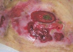
Case study: Peristomal pyoderma gangrenosum

As a wound care specialist, you have learned about many skin conditions, some so unusual and rare that you probably thought you would never observe them. I’ve been a nurse for 38 years, with the last 10 years in wound care, and that’s certainly what I thought. But I was wrong. Let me tell you about my challenging patient with an unusual skin condition.

A perplexing patient (more…)

Read More

2015 Journal: Best of the Best Vol. 4 No. 5

Wound Care Advisor Journal Best of the Best vol.4 no5

Evolution of the deep tissue injury or a declining pressure ulcer?

A declining pressure ulcer decreases the quality of life for patients and places providers at risk for regulatory citations and litigation. But it’s important for clinicians to determine whether the first appearance of skin injury is truly a stage I or II pressure ulcer or if it’s a deep tissue injury (DTI), a unique staging category for a pressure ulcer. Otherwise, a clinician might think a pressure ulcer is getting worse instead of the change being the normal progression of a pressure ulcer that is presenting as a DTI.

Read more

“Best of the Best” three-peat

By: Donna Sardina, RN, MHA, WCC, CWCMS What do the Los Angeles Lakers, Green Bay Packers, Montreal Canadiens, and New York Yankees have in common? All three have “three-peated”, meaning they have won three consecutive championships. This year, we at Wound Care Advisor, the official journal of the National Alliance of Wound Care and Ostomy (NAWCO), mark our own three-peat—our…

Clinical Notes

Mild compression diabetic socks safe and effective for lower extremity edema Diabetic socks with mild compression can reduce lower extremity edema in patients with diabetes without adversely affecting arterial circulation, according to a randomized control trial presented at the American Diabetes Association 75th Scientific Sessions Conference.

Clinician Resources: NPUAP, Pressure Ulcer Treatment, NIOSH

The resources below will help you address issues in your practice. NPUAP position statement on hand check for bottoming out Use of the hand check to determine “bottoming out” of support systems should be limited to static air overlay mattresses, according to a position statement from the National Pressure Ulcer Advisory Panel (NPUAP).

Creating effective education programs on a shoestring budget

By Jennifer Oakley, BS, RN, WCC, DWC, OMS It’s time again for annual staff education, and you, the certified wound clinician, need to teach the staff at your organization. You dream of staff entering a state-of-the-art classroom with computers at each station, mannequins, wound anatomy models, and enough products for each student to do hands-on demonstrations. But when you open…

Evolution of the deep tissue injury or a declining pressure ulcer?

 By Jeri Lundgren, BSN, RN, PHN, CWS, CWCN A declining pressure ulcer decreases the quality of life for patients and places providers at risk for regulatory citations and litigation. But it’s important for clinicians to determine whether the first appearance of skin injury is truly a stage I or II pressure ulcer or if it’s a deep tissue injury (DTI),…

Get the ‘SKINNI’ on reducing pressure ulcers

By Cindy Barefield, BSN, RN-BC, CWOCN Like many hospitals, Houston Methodist San Jacinto Hospital uses national benchmarks such as the National Database of Nursing Quality Indicators (NDNQI®) to measure quality outcomes. Based on benchmark reports that showed an increased trend of pressure ulcers in critically ill patients in our hospital, the clinical nurses in our Critical Care Shared Governance Unit-Based…

Helping patients overcome ostomy challenges

By Beth Hoffmire Heideman, MSN, RN No one wants an ostomy, but sometimes it’s required to save a patient’s life. As ostomy specialists, our role is to assess and intervene for patients with a stoma or an ostomy to enhance their quality of life. We play an active role in helping patients perform self-care for their ostomy and adjust to…

how to assess wound exudate

How to assess wound exudate

By Nancy Morgan, RN, BSN, MBA, WOC, WCC, DWC, OMS Each issue, Apple Bites brings you a tool you can apply in your daily practice. Exudate (drainage), a liquid produced by the body in response to tissue damage, is present in wounds as they heal. It consists of fluid that has leaked out of blood vessels and closely resembles blood…

Medical gauze 101

By Nancy Morgan, RN, BSN, MBA, WOC, WCC, DWC, OMS Each issue, Apple Bites brings you a tool you can apply in your daily practice. Medical gauze, a bleached white cloth or fabric used in bandages, dressings, and surgical sponges, is the most widely used wound care dressing. Commonly known as “4×4s,” gauze is made from fibers of cotton, rayon,…

Palliative wound care: Part 1

By Gail Rogers Hebert, MS, RN, CWCN, WCC, DWC, OMS, LNHA The World Health Organization defines palliative care as “an approach that improves the quality of life of patients and their families facing the problem associated with life-threatening illness, through the prevention and relief of suffering by means of early identification and impeccable assessment and treatment of pain and other…

Providing evidence-based care for patients with lower-extremity cellulitis

By Darlene Hanson, PhD, RN; Diane Langemo, PhD, RN, FAAN; Patricia Thompson, MS, RN; Julie Anderson, PhD, RN; and Keith Swanson, MD Cellulitis is an acute, painful, and potentially serious spreading bacterial skin infection that affects mainly the subcutaneous and dermal layers. Usually of an acute onset, it’s marked by redness, warmth, swelling, and tenderness. Borders of the affected skin…

2015 Journal: Best of the Best Vol. 4 No. 5
Read More

Providing evidence-based care for patients with lower-extremity cellulitis

By Darlene Hanson, PhD, RN; Diane Langemo, PhD, RN, FAAN; Patricia Thompson, MS, RN; Julie Anderson, PhD, RN; and Keith Swanson, MD

Cellulitis is an acute, painful, and potentially serious spreading bacterial skin infection that affects mainly the subcutaneous and dermal layers. Usually of an acute onset, it’s marked by redness, warmth, swelling, and tenderness. Borders of the affected skin are characteristically irregular. Although cellulitis may occur in many body areas, this article discusses the most common location—the lower limb. (more…)

Read More

2015 Journal: May – June Vol. 4 No. 3

Wound Care Advisor Journal Vol4 No3

Get the ‘SKINNI’ on reducing pressure ulcers

Like many hospitals, Houston Methodist San Jacinto Hospital uses national benchmarks such as the National Database of Nursing Quality Indicators (NDNQI®) to measure quality outcomes. Based on benchmark reports that showed an increased trend of pressure ulcers in critically ill patients in our hospital, the clinical nurses in our Critical Care Shared Governance Unit-Based Council (CCSGUBC) identified an improvement opportunity.

Read more

A collaborative approach to wound care and lymphedema therapy: Part 1

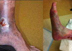
By Erin Fazzari, MPT, CLT, CWS, DWC Have you seen legs like those shown in the images below in your practice? These images show lymphedema and venous stasis ulcers, illustrating the importance of collaboration between clinicians in two disciplines: lymphedema and wound care.

Ankle-brachial index: A dirty word?

Donna Sardina, RN, MHA, WCC, CWCMS, DWC, OMS Silence, roving eyes, fidgeting, excuses, a quick subject change—these are typical responses from healthcare clinicians when asked, “What’s the patient’s ankle-brachial index?” You’d think someone had just uttered a dirty word. The ankle-brachial index (ABI) is a key component of the lower-extremity vascular exam, recommended and in some cases mandated by numerous…

Clinical Notes: diabetes, LMW heparin, dressings, lymphedema

Factors affecting medication adherence in patients with diabetes identified Factors associated with better adherence to antidiabetic medications taken by patients with diabetes include older age, male sex, higher education, higher income, use of mail-order vs. retail pharmacies, primary care vs. nonendocrinology specialist prescribers, higher daily total pill burden, and lower out-of-pocket costs.

Clinician Resources: Nutrition, Workplace Violence, Pressure Injuries

Learn about resources useful to your practice. Nutrition and pressure ulcers Advances in Skin & Wound Care has published “The role of nutrition for pressure ulcer management: National Pressure Ulcer Advisory Panel, European Pressure Ulcer Advisory Panel, and Pan Pacific Pressure Injury Alliance White Paper.” The white paper includes evidence-based nutrition strategies for preventing and managing pressure ulcers.

Creating high-performance interprofessional teams

By Terry Eggenberger, PhD, RN, CNE, CNL; Rose O. Sherman, EdD, RN, NEA-BC, FAAN; and Kathryn Keller, PhD, RN Kate Summer, a wound care clinician in a urban hospital, is leading an initiative to reduce pressure ulcers. She knows from experience that more effective communication and collaborative planning by the interdisciplinary team managing these patients is crucial for reducing pressure…

Get the ‘SKINNI’ on reducing pressure ulcers

By Cindy Barefield, BSN, RN-BC, CWOCN Like many hospitals, Houston Methodist San Jacinto Hospital uses national benchmarks such as the National Database of Nursing Quality Indicators (NDNQI®) to measure quality outcomes. Based on benchmark reports that showed an increased trend of pressure ulcers in critically ill patients in our hospital, the clinical nurses in our Critical Care Shared Governance Unit-Based…

Helping patients overcome ostomy challenges

By Beth Hoffmire Heideman, MSN, RN No one wants an ostomy, but sometimes it’s required to save a patient’s life. As ostomy specialists, our role is to assess and intervene for patients with a stoma or an ostomy to enhance their quality of life. We play an active role in helping patients perform self-care for their ostomy and adjust to…

Helping patients with lower-extremity disease benefit from exercise

By Jeri Lundgren, BSN, RN, PHN, CWS, CWCN Research has shown that exercise can help ease symptoms in patients with arterial insufficiency, venous insufficiency, neuropathic disease, or a combination of these conditions. Here’s what you need to know to ensure your patients reap the most benefits from exercise.

Moldable ostomy barrier rings and strips

By Nancy Morgan, RN, BSN, MBA, WOC, WCC, DWC, OMS Each issue, Apple Bites brings you a tool you can apply in your daily practice. Here’s a brief overview on moldable, bendable, and stretchable adhesive rings and strips used to improve the seal around a stoma. Benefits Adhesive rings and strips can be an alternative to stoma paste for filling…

Providing evidence-based care for patients with lower-extremity cellulitis

By Darlene Hanson, PhD, RN; Diane Langemo, PhD, RN, FAAN; Patricia Thompson, MS, RN; Julie Anderson, PhD, RN; and Keith Swanson, MD Cellulitis is an acute, painful, and potentially serious spreading bacterial skin infection that affects mainly the subcutaneous and dermal layers. Usually of an acute onset, it’s marked by redness, warmth, swelling, and tenderness. Borders of the affected skin…

2015 Journal: May – June Vol. 4 No. 3
Click here to access the digital edition
Read More

Palliative wound care: Part 2

This approach brings patient-centered care to life.

 By Gail Rogers Hebert, MS, RN, CWCN, WCC, DWC, OMS, LNHA

Editor’s note: This article is the second in a two-part series on palliative wound care. For the first part, click here.

By preventing and relieving suffering, palliative care improves the quality of life for patients facing problems associated with life-threatening illness. This care approach emphasizes early identification, impeccable assessment, and treatment of pain and other issues—physical, psychosocial, and spiritual. (more…)

Read More

Evolution of the deep tissue injury or a declining pressure ulcer?

 By Jeri Lundgren, BSN, RN, PHN, CWS, CWCN

A declining pressure ulcer decreases the quality of life for patients and places providers at risk for regulatory citations and litigation. But it’s important for clinicians to determine whether the first appearance of skin injury is truly a stage I or II pressure ulcer or if it’s a deep tissue injury (DTI), a unique staging category for a pressure ulcer. Otherwise, a clinician might think a pressure ulcer is getting worse instead of the change being the normal progression of a pressure ulcer that is presenting as a DTI. (more…)

Read More
1 2 3 4