By Darlene Hanson, PhD, RN; Diane Langemo, PhD, RN, FAAN; Patricia Thompson, MS, RN; Julie Anderson, PhD, RN; and Keith Swanson, MD
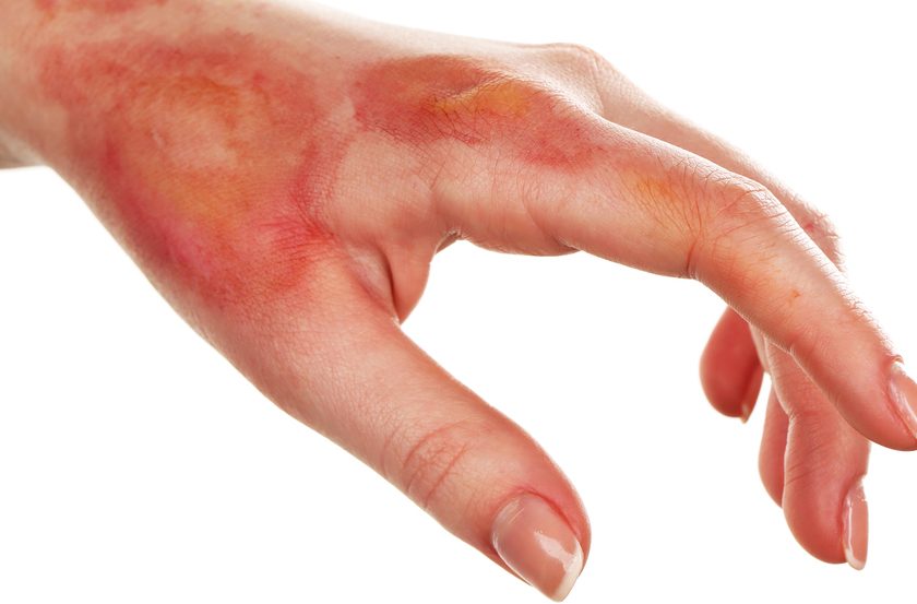
Cellulitis is an acute, painful, and potentially serious spreading bacterial skin infection that affects mainly the subcutaneous and dermal layers. Usually of an acute onset, it’s marked by redness, warmth, swelling, and tenderness. Borders of the affected skin are characteristically irregular. Although cellulitis may occur in many body areas, this article discusses the most common location—the lower limb.
In cellulitis, bacteria enter through an opening in the skin caused by a bite, an ulcer, a body piercing, or other discontinuity. The most common bacteria are Streptococcus pyogenes and Staphylococcus aureus, which are indigenous to the skin. The body reacts to these microbes as foreign, leading to presenting signs and symptoms. On assessment, clinicians may notice a recent insect bite, surgical incision, or trauma to the leg.
Cellulitis and cutaneous abscesses combined cause nearly 600,000 hospital admissions annually in the United States—an increase of 65% since 1999. Cellulitis and other soft-tissue infections account for up to 10% of hospital admissions. Incidence of cellulitis ranges from 0.2 in 1,000 person-years to 24.6 in 1,000 person-years in different populations.
In 2006, about 14.5 million cases of cellulitis occurred, incurring costs of approximately $3.7 billion overall. Costs may rise when the condition is misdiagnosed or when antibiotics are used inappropriately, as this may prolong treatment or predispose patients to complications.
What the literature shows
In 2012, Lipsky and colleagues completed a prospective multicenter study of patients with soft-tissue infections to explore the epidemiology, clinical presentation, treatment, and clinical outcomes. Of the 1,033 subjects, 26.9% had cellulitis and the same percentage had diabetic foot infections. In contrast, surgical-site infections affected 16.7% and deep soft-tissue abscesses affected 13.6%. The lower leg was the most common cellulitis site (49.6%). Pain was rated as moderate to severe in 73% (n = 203) of patients. Overall, patients with cellulitis had more severe erythema and local warmth than those with other soft-tissue infections. However, abscess, induration, tenderness, and pain were more common and more severe in patients with deep soft-tissue abscess. Leg warmth was absent in only 10 of the 278 cellulitis patients.
Comorbidities most often accompanying cellulitis included diabetes, peripheral vascular disease, chronic lung disease, and renal insufficiency. Treatment included initial I.V. vancomycin in 60% of patients, followed by penicillins, beta-lactamase inhibitors, and cephalosporins. For patients hospitalized with cellulitis, the mean stay was 7.1 days (range, 5.8 to 8.1 days).
A 2010 study by Kilburn and colleagues found 25 randomized controlled trials related to cellulitis. The review noted that macrolides reportedly were more effective than penicillins in treating cellulitis and oral antibiotics were more effective than I.V. antibiotics. But due to lack of research-supported findings, reviewers couldn’t give specific recommendations for cellulitis treatment; further study is needed to determine the best treatment.
A retrospective epidemiologic and outcomes study by Zervos and colleagues (in 2012) assessed the origin of complicated and soft-tissue infections and the appropriateness of initial antibiotic therapy in hospital patients. In the sample of 1,096 patients, the most common soft-tissue infections were cellulitis and abscess, usually community acquired. S. aureus was the most common culture-positive skin infection; 74% of these infections were methicillin-resistant. More work needs to be done to examine the impact of skin infections and use of appropriate initial therapy for such infections.
Risk factors
Cellulitis is common in patients with circulatory problems of the legs, particularly those with venous disease. Anyone who sustains leg trauma, an insect bite, or a surgical wound is at risk. People who are overweight or have leg ulcers or lymphedema are at higher risk. Lymphedema especially increases cellulitis risk because the lymphatic pathways transport immune cells to fight infection; if these pathways are blocked, cellulitis can readily occur.
Cellulitis isn’t contagious because it’s an infection of the dermis and subcutaneous tissues, which act as a protective layer over the infected tissues. Rarely, it can lead to a deeper, more serious skin infection, such as necrotizing fasciitis.
Diagnosis, staging, and classification
Clinical Resource Efficiency Support Team (CREST) guidelines aid diagnosis. Cellulitis ranges from class I to class IV, with IV being the most severe.
- Class I: Patients lack systemic signs or symptoms.
- Class II: Patients have comorbid conditions that affect recovery.
- Class III: Patients have accompanying limb-threatening conditions or confusion, tachycardia, or other unstable conditions.
- Class IV: Patients have severe, life-threatening infections or septicemia. (See Classifying cellulitis.)
Differentiating cellulitis from similar conditions
Cellulitis is diagnosed definitively based on classic symptoms, which include a unilateral hot, erythematous, nonblanching redness that persists with limb elevation. Skin may be dry and flaking. Commonly, subcutaneous tissue is tender; in severe cellulitis, crepitations may occur.
Differentiating cellulitis from other conditions may prove challenging. One study with a sample of 635 patients who’d been diagnosed with cellulitis found only 425 (67%) actually had the condition. Disorders that can mimic cellulitis include eczema, tinea pedis, and other chronic conditions such as erysipelas. Lipodermatosclerosis also may be mistaken for cellulitis.
Unlike cellulitis, venous eczema can cause a range of manifestations, such as bilateral symptoms, itching, hemosiderin deposits, and edema. Suspect venous eczema, not cellulitis, in a patient with reddened leg skin, chronic venous disease or an ulcer, and a history of appropriate antibiotics with no improvement.
Dependent rubor from peripheral vascular disease also may resemble cellulitis. But in this condition, further assessment reveals short-distance claudication or “rest” pain, lack of hair growth on the lower limb, and redness that completely disappears on elevation.
Assessing for induration
If you suspect cellulitis, assess for induration—a hardened mass or formation with defined edges, with slight swelling and firmness at the edges or border between normal skin and skin affected by cellulitis. The Bates-Jensen Wound Assessment Tool recommends assessing induration by gently attempting to pinch the affected area; with induration, you won’t be able to pinch the tissue. Use a measuring tool to document how far induration extends. Wound care clinicians typically outline the indurated area from visit to visit to determine if induration has increased or decreased.
Treatment
Guidelines for cellulitis treatment hinge on severity. A triple approach using I.V. antibiotics, I.V. fluids, and pain management is recommended. Light compression is suggested if the ankle-brachial index (ABI) is adequate, but use caution during an acute cellulitis episode. Consider limb elevation and analgesics for comfort.
Treatment should be prompt to help prevent complications. Using the HAMMMER acronym can help you remember the essential elements of treatment. (See Cellulitis treatment and HAMMMER interventions.)
Antibiotics
Clinicians typically prescribe a 14-day course of antibiotics (unless contraindicated) if they’re unsure whether inflammation stems from infection. Advise patients to contact their primary care practitioner if they don’t notice a response to therapy within 3 days. Antibiotics are effective in about 90% of cases. If the affected area is quite small and cellulitis isn’t severe, it may clear without antibiotics; if exudate is more than minimal, the patient usually needs antibiotics.
Empirical treatment with semisynthetic penicillin, first-or second-generation cephalosporins, macrolides, or clindamycin is advised, primarily because of the increasing incidence of methicillin-resistant S. aureus (MRSA) or erythromycin-resistant S. pyogenes. When cellulitis surrounds an abscess formation with MRSA, about half of the infections resist clindamycin. Of the S. pyogenes cases resistant to macrolides, about 99.5% are susceptible to clindamycin and 100% to penicillin. If the condition doesn’t improve, symptoms are extensive, or the patient has a high temperature, hospitalization and I.V. antibiotics may be warranted.
I.V. fluids and hydration
As with any systemic infection, I.V. fluids are indicated, as the infection can significantly increase insensible water loss, in turn causing dehydration and possibly multisystemic failure.
Compression
In the past, studies recommended against using compression, assuming it could spread bacteremia. Current best practice includes light compression therapy used cautiously. (Acute infections that lead to swelling can cause higher tissue pressures than normal and compression could further compromise the limb.) Teach the patient how to apply and care for the compression hose. Before considering compression in any form, perform a vascular assessment, including ABI measurement. (See Cellulitis: A case study.)
Pain management and skin comfort
Assess the patient’s pain level and provide pain management as needed. Nonsteroidal anti-inflammatory drugs hasten healing when combined with antibiot-ics. Moisturizing the limb can reduce skin dryness and flaking and ease discomfort.
Limb elevation
Elevating the affected leg above heart level is a key intervention for cellulitis. Raise the ankle higher than the knee, the knee higher than the hip, and the entire leg higher than heart level. Continue elevation for the first 24 to 48 hours while I.V. antibiotics are infusing.
Monitoring for complications
Measure the patient’s temperature on an ongoing basis. Expect to obtain blood cultures as a standard of care. For complex patients with peripheral arterial disease, assess for complications, such as gangrene and poorly healing wounds.
If cellulitis doesn’t respond to ordinary treatment, suspect complications, such as septicemia. This condition arises when bacteria spread to the lymph system and bloodstream. Rarely, the infection may spread to deeper fascial tissues (resulting in necrotizing fasciitis) or to the bone (causing osteomyelitis). Signs and symptoms of systemic infection include chills, sweating, fatigue, general malaise, muscle ache, and a sensation of heat. These require prompt attention.
Recurrent cellulitis can damage the lymphatic drainage system of the affected limb, causing lymphangitis, chronic lymphedema, or both. Also, abscesses may form if the infection becomes highly localized in a small area.
Innovations in therapy
In England, a nurse-led “Red Legs” service has been established to help meet the needs of patients with conditions that can be misconstrued as cellulitis. A team of healthcare professionals established integrated care pathways for cellulitis diagnosis and treatment. Results were promising and included a significant cost savings. Another group of British researchers reported on the effectiveness of training caregivers about cellulitis using simulation methods. In a 2011 simulation study by Unsworth and colleagues, nurses who participated in patient simulation scenarios had a 45% increase in confidence levels regarding diagnosing and managing cellulitis and recognizing patient deterioration. Further research is needed so healthcare professionals can provide cost-effective, evidence-based treatment for the many individuals affected by cellulitis.
Selected references
Beasley A. Management of patients with cellulitis of the lower limb. Nurs Stand. 2011;26(11):50-5.
Clinical Resource Efficiency Support Team (CREST). Guidelines on the Management of Cellulitis in Adults. June 2005. www.acutemed.co.uk/docs/Cellulitis guidelines, CREST, 05.pdf
Dalal A, Eskin-Shwartz M, Mimouni D, et al. Interventions for the prevention of recurrent erysipelas and cellulitis. Cochrane Database Syst Rev. 2012:4:CD009758.
Elwell R. Developing a nurse-led integrated ‘red legs’ service. Br J Community Nurs. 2014;19(1):12-9.
Harris C, Bates-Jensen B, Parslow N, et al. Bates-Jensen wound assessment tool: pictorial guide validation project. J Wound Ostomy Continence Nurs. 2010;37(3):253-9.
Jenkins TC, Knepper BC, Sabel AL, et al. Decreased antibiotic utilization after implementation of a guideline for inpatient cellulitis and cutaneous abscess. Arch Intern Med. 2011;171(12):1072-9.
Kilburn SA, Featherstone P, Higgins B, Brindle R. Interventions for cellulitis and erysipelas. Cochrane Database Syst Rev. 2010 June 16;(6):CD004299.
Levell NJ, Wingfield CG, Garioch JJ. Severe lower limb cellulitis is best diagnosed by dermatologists and managed with shared care between primary and secondary care. Br J Dermat. 2011;164(6):1326-8.
Lipsky BA, Moran GJ, Napolitano LM, et al. A prospective, multicenter, observational study of complicated skin and soft tissue infections in hospitalized patients: clinical characteristics, medical treatment, and outcomes. BMC Infect Dis. 2012;12:227.
Stevens DL, Bisno AL, Chambers HF, et al. Practice guidelines for the diagnosis and management of skin and soft tissue infections: 2014 update by the Infectious Disease Society of America. Clin Infect Dis. 2014;59(2):147-59.
Unsworth J, Tuffnell C, Platt A. Safer care at home: use of simulation training to improve standards. Br J Community Nurs. 2011;16(7):334-9.
Wingfield C. Diagnosing and managing lower limb cellulitis. Nurs Times. 2012;108(27):18-21.
Wound, Ostomy and Continence Nurses Society (WOCN). Guideline for management of wounds in patients with lower-extremity venous disease. Mt. Laurel, NJ: Author; 2011.
Zervos MJ, Freeman K, Vo L, et al. Epidemiology and outcomes of complicated skin and soft tissue infections in hospitalized patients. J Clin Microbiol. 2012;50(2):238-45.
Darlene Hanson is a clinical associate professor at the University of North Dakota (UND) College of Nursing and Professional Disciplines in Grand Forks. Diane Langemo is a professor emeritus, Patricia Thompson is a clinical associate professor, and Julie Anderson is professor and acting director of library sciences at UND. Keith Swanson is a physician specializing in internal medicine and vascular medicine at the Altru Health System in Grand Forks.
DISCLAIMER: All clinical recommendations are intended to assist with determining the appropriate wound therapy for the patient. Responsibility for final decisions and actions related to care of specific patients shall remain the obligation of the institution, its staff, and the patients’ attending physicians. Nothing in this information shall be deemed to constitute the providing of medical care or the diagnosis of any medical condition. Individuals should contact their healthcare providers for medical-related information.