One Doctor Exploring Wound Care on Earth and in Space

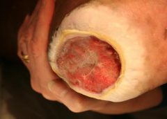
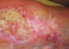
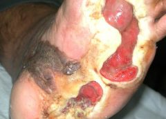
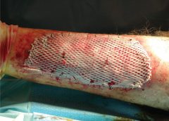
In laboratories all across the globe, scientists are uncovering new and exciting breakthroughs in the realm of wound healing.

For instance, a team out of Texas is blinding bacteria to prevent their spread. Meanwhile, a collective of doctors from the U.K. recently developed some intriguing new vacuum tech to treat chronic ulcers. There’s even been research into drug treatments, like how opioids may actually prevent proper wound care.

Each team has taken a different approach or tackled a unique situation or medical ailment, and that ensures a more well-rounded coverage that helps a larger pool of patients. However, few scientists have a more grand scope than Ronke Olabisi, a professor of biomedical engineering at Rutgers University.

Reaching for the stars

As the university explained in a recent press release, Olabisi is hard at work on several projects aimed at improving wound healing both on earth and during manned space missions. During space travel, especially as astronauts spend months at a time in stations, the lack of gravity has a huge impact on the human body. Muscle and bones will actually start to deteriorate, and tissues will lose much of their elasticity. Olabisi’s main goal is to study in-depth why this occurs and how to fix, and she believes she can apply much of the same knowledge to wound care on Earth.

Read more at Advanced Tissue

Read More

Accuracy of the Ankle-brachial Index in the Assessment of Arterial Perfusion of Heel Pressure Injuries

Abstract: Background. The evaluation and treatment of heel pressure injuries are a significant and expensive sequela of the aging population. Although the workup of patients with lower extremity tissue loss usually involves an assessment of the arterial blood flow by means of noninvasive vascular testing, the results may be misleading in patients with heel pressure injuries when the ankle-brachial index (ABI) does not provide direct information about perfusion of the rearfoot. The objective of this retrospective, observational investigation was to determine if noninvasive vascular testing provides accurate and reliable results in patients with heel pressure injuries. (more…)

Read More

Wound Photography – How it Benefits Clinical Documentation

wound photography

Accurate assessment and documentation of wounds is essential for developing a comprehensive plan of care. Photography now plays a key role in wound care. The use of digital photography has enhanced the reliability and accuracy of wound documentation. Though a wound assessment in patient files includes details such as location, depth, odor, condition of surrounding tissue and other details, a visual record can be worth even more.

Digital photography is becoming a more prevalent documentation tool. According to an article published in McKnight’s, forensic nursing experts recommend using photographs to document injury. The photos show both how an injury occurred and how it is healing.

The National Pressure Ulcer Advisory Panel (NPUAP) also supports photography as a more accurate means for assessment of wound dimensions and wound base over time.

A visual confirmation to the written record, these images:

  • Facilitate better diagnosis
  • Enhance clinical documentation
  • Help to monitor the progress of wound healing
  • Help prevent litigation in wound management
  • Allow inter-disciplinary communication among the wound care team

Read more at Wound Wizard

Read More

Crawford bags FDA clearance for wound dressing that will ‘save limbs’

Crawford Healthcare, one of the biggest makers of advanced wound-care products in the UK, has won clearance from US regulators for a medical dressing that it says will “save limbs”.

The product, called KerraCel AG, soaks up fluid and bacteria from nasty, oozing wounds and locks it away as a gel. It is also the only dressing of its kind to contain silver at a special concentration to kill all bacteria – even those resistant to antibiotics – that prevent chronic wounds, such as diabetic ulcers and pressure sores, from healing.

Read more at The Telegraph

Read More

Management of Patients With Venous Leg Ulcers

It is well documented that the prevalence of venous leg ulcers (VLUs) is increasing, coinciding with an ageing population. Accurate global prevalence of VLUs is difficult to estimate due to the range of methodologies used in studies and accuracy of reporting.1 Venous ulceration is the most common type of leg ulceration and a significant clinical problem, affecting approximately 1% of the population and 3% of people over 80 years of age2 in westernised countries. Moreover, the global prevalence of VLUs is predicted to escalate dramatically, as people are living longer, often with multiple comorbidities. Recent figures on the prevalence of VLUs are based on a small number of studies, conducted in Western countries, and the evidence is weak. However, it is estimated that 93% of VLUs will heal in 12 months, and 7% remain unhealed after five years.3 Furthermore, the recurrence rate within 3 months after wound closure is as high as 70%.4-6 Thus, cost-effective adjunct evidence-based treatment strategies and services are needed to help prevent these ulcers, facilitate healing when they occur and prevent recurrence.

The impact of a VLU represents social, personal, financial and psychological costs on the individual and further economic drain on the health-care system. This brings the challenge of providing a standardised leg ulcer service which delivers evidence-based treatment for the patient and their ulcer. It is recognised there are variations in practice and barriers preventing the implementation of best practice. There are patients not receiving appropriate and timely treatment in the initial development of VLUs, effective management of their VLU and preventing recurrence once the VLU has healed.

Health-care professionals (HCPs) and organisations must have confidence in the development process of clinical practice guidelines and have ownership of these guidelines to ensure those of the highest quality guide their practice. These systematic judgments can assist in policy development, and decision making, improve communication, reduce errors and improve patient outcomes.

Read more at Journal of Wound Care

Read More

Reduction of 50% in Diabetic Foot Ulcers With Stem Cells

Diabetic Foot Ulcers

MUNICH — Local injection of mesenchymal stem cells derived from autologous bone marrow shows promise in healing recalcitrant neuropathic diabetic foot ulcers, a novel study from Egypt shows.

Presenting the results at the European Association for the Study of Diabetes (EASD) 2016 Annual Meeting, Ahmed Albehairy, MD, from Mansoura University, Egypt, said: “In patients who received the mesenchymal stem cells, ulcer reduction was found to be significantly higher compared with patients on conventional treatment after both 6 weeks and 12 weeks of follow-up. This is despite the fact that initial ulcer size was larger in the stem-cell–treated group.” (more…)

Read More

Better Skin Grafts – take only one layer

skin grafts take one layer

Research shows that a skin-graft harvesting system aids chronic wound recovery and reduces care costs by accelerating the healing process.

More than six million cases of chronic wounds cost $20 billion each year in the United States. Diabetic ulcers, pressure sores, surgical site wounds, and traumatic injuries to high-risk patients account for most wounds that won’t heal. (more…)

Read More