By Erin Fazzari, MPT, CLT, CWS, DWC
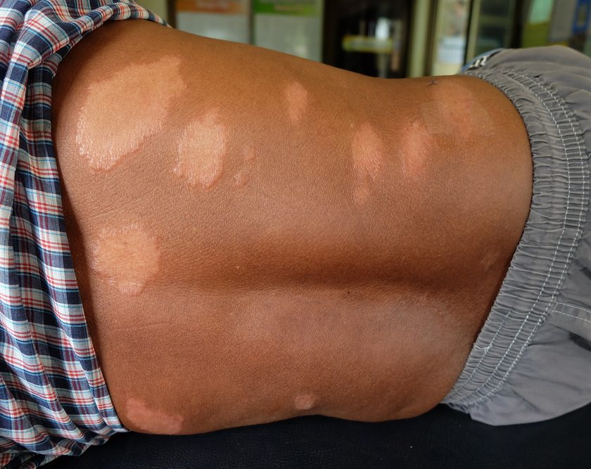
Have you seen legs like those shown in the images below in your practice? These images show lymphedema and venous stasis ulcers, illustrating the importance of collaboration between clinicians in two disciplines: lymphedema and wound care.
My experience
Over the last 12 years as a physical therapist specializing in lymphedema therapy and wound care, I’ve had the opportunity to treat many patients with wounds in multiple settings. I’ve also had the opportunity to collaborate with medical professionals in multidisciplinary treatment centers where lymphedema therapists and wound care clinicians act as a team. Through this experience—and through review of the literature—I’ve learned that such a team has improved patient outcomes.
To help the team reap maximal benefits, I’d like to share information related to lymphedema, its management, and how collaboration in multidisciplinary treatment centers can enhance outcomes.
In Part 1 of this two-part series, I discuss pathophysiology related to wounds and lymphedema and begin the discussion of collaboration.
The basics
To understand the role of lymphedema therapy as it relates to wound care, it’s first necessary to take a step back and define both chronic wound and lymphedema. A chronic wound is a wound that doesn’t heal in an orderly set of stages and in the predictable amount of time that most wounds do. Delayed healing may result from a variety of underlying factors, such as poor systemic immune function, malnourishment, chemotherapeutic agents, high bioburden, repetitive mechanical trauma, and cytotoxic agents.
Lymphedema is a condition of localized fluid retention and tissue swelling characterized by high-protein edema caused by a compromised lymphatic system. All exterior regions of the body (for example, face, neck, torso, extremities, and genitals) can be affected. Common causes of lymphedema and accompanying diseases that can contribute to lymphedema include heredity, filariasis, trauma, surgeries, lymph node dissections, radiation therapy, malignancy, obesity, diabetes, chronic heart failure, dependent mobility and, of course, venous disease—the principal culprit of our wound for discussion, the venous stasis ulcer.
The venous stasis ulcer is one major debilitating result of advanced venous disease. Venous ulceration is the most common cause of lower-extremity ulcer, accounting for half of these ulcers and affecting 1% to 2% of the U.S. population, with 3% to 5% of patients older than age 65.
Venous stasis ulcers and lymphedema
So how are the venous ulcer and lymphedema related? The venous and lymphatic systems are closely intertwined. When explaining the systems to patients, I often refer to the lymphatics as the sewer system of the venous system.
Most wound care clinicians are familiar with the pathophysiology that results in venous disease and the cascade of events that leads to a venous ulcer. Many clinicians, however, aren’t as familiar with the role of the lymphatics in this process.
Under optimal circumstances, the venous system is responsible for the removal of 90% of interstitial fluid at the capillary level. The remaining 10% is the responsibility of the lymphatic system. However, the lymphatics have a built-in safety net to manage excess interstitial fluid that occurs when the veins function inefficiently or ineffectively. Venous reflux may be present, but edema in the tissue isn’t yet visible when the lymphatics are able to manage the load. Edema in the lower extremities, as well as other areas of the body, is visible only when both the veins and lymphatics are no longer capable of managing the load.
The lymphatics are also responsible for the removal of large macromolecules from the interstitial space, including proteins that are unable to diffuse back into the venous system at the capillary level. A venous stasis ulcer occurs when the increase in protein concentrations in the tissue results in chronic inflammation and infiltration of white blood cells and fibroblasts. This leads to fibrosis of the edematous tissue, dilation and insufficiency of lymphatic tissue, and damage to endothelial cells, further reducing lymphatic flow and enhancing the destructive process.
The body’s physiologic responses illustrate the close anatomic and physiologic connection between the two systems. Consequently, it should be a priority for us, as clinicians, to address both the lymphatic and venous systems when edema is detectable in the tissue.
Worldwide impact
The anatomy and physiology of these systems have a huge impact on our patient population. Each year in North America, 5 to 7 million chronic wounds occur. Lower-extremity venous stasis ulcer is the most common of these, with an incidence of 2.5 million. In the United States alone, chronic leg wounds account for 2 million lost workdays per year.
When it comes to lymphedema, 1 in 30 people worldwide are estimated to be afflicted with this debilitating disease, not including those suffering from venous disease.
World organizations have begun to recognize the importance of addressing lymphedema and wound care collaboratively. (See World action on lymphedema and wound care.) As noted earlier, anatomy and physiology don’t separate the venous and lymphatic systems, so wound care and lymphedema clinicians need to work collaboratively to help patients.
Common goals
A good place to start collaborating is to understand that the disciplines of lymphedema therapy and wound care have many common goals, including:
- reducing and stabilizing edema
- achieving ulcer healing
- preventing recurrence
- preventing infection
- maximizing tissue healing.
Multidisciplinary teams are the wave of the future of health care. Consider how a team approach with lymphedema and wound care professionals would enhance your practice, and watch for Part 2 of this series, which will further address common goals, review the gold standard of management (complex decongestive therapy), and illustrate how collaboration in multidisciplinary treatment centers can enhance patient outcomes.
Selected references
Ditzler K. Collaborating lymphedema and wound care [lecture]. Penn Medicine at Radnor, Radnor, PA; 2014.
Foldi M, Foldi E. Foldi’s Textbook of Lymphology: For Physicians and Lymphedema Therapists. 3rd ed. Munich, Germany: Urban & Fischer, 2012.
Law K. Addressing the Whole, Not Just the Hole: A Collaborative Approach for Patient Success [Lecture]. Penn Medicine at Radnor, Radnor, PA; 2014.
MacDonald J, Asiedu K. WAWLC: World alliance for wound and lymphedema care. Wounds. 2010; 22(3):55-9.
MacDonald J, Geyer, MJ, eds. Wound and lymphoedema management. World Health Organization: 2010 http://whqlibdoc.who.int/publications/2010/9789241599139_eng.pdf
Macdonald JM, Sims N, Mayrovitz HN. Lymphedema, lipedema, and the open wound: the role of compression therapy. Surg Clin North Am. 2003;83(3):639-58.
Norton S. CDT theoretical review course for the certified lymphedema therapist. Norton School of Lymphatic Therapy; 2005.
Pawel D, Franek A, Kolank M. A comparative clinical study on five types of compression therapy in patients with venous leg ulcers. Int J Med Sci. 2013 Dec;11(1):34-43.
Sen CK, Gordillo GM, Roy S, et al. Human skin wounds: a major and snowballing threat to public health and the economy. Wound Repair Regen. 2009 Nov-Dec;17:763-7.
Erin Fazzari is a physical therapist at Good Shepherd Penn Partners: Penn Therapy and Fitness, in Philadelphia, Pennsylvania.