Gene Therapy for Non-Healing Diabetic Foot Ulcers Starts Phase III Trial

Diabetic Foot Ulcers

Safety and Efficacy Study of VM202 in the Treatment of Chronic Non-Healing Foot Ulcers. This study will assess the safety and efficacy of using gene therapy via intramuscular injections of the calf for patients with chronic non-healing foot ulcers.

The first patient has been dosed in a Phase III trial assessing ViroMed’s VM202, the first pivotal study of a gene therapy indicated for patients with nonhealing diabetic foot ulcers (NHU) and concomitant peripheral artery disease (PAD).

The Phase III trial (NCT02563522) is a double-blind, placebo-controlled, multicenter study designed to evaluate VM202 for safety and efficacy in 300 adults with a diabetic foot ulcer and concomitant PAD. Two hundred patients will be randomized to VM202 and the other 100 to placebo, ViroMed’s U.S. division VM BioPharma said yesterday. (more…)

Read More

Wounds that won’t heal can be devastating

diabetic foot ulcer

Carol Emanuele beat cancer. But for the last two years, the Philadelphia woman has been fighting her toughest battle yet. She has an open wound on the bottom of her foot that leaves her unable to walk and prone to deadly infection.

In an effort to treat her diabetic wound, doctors at a clinic in Northeast Philadelphia have prescribed a dizzying array of treatments. Freeze-dried placenta. Penis foreskin cells. High doses of pressurized oxygen. And those are just a few of the treatment options patients face.

“I do everything, but nothing seems to work,” said Emanuele, 59, who survived stage 4 melanoma in her 30s. “I beat cancer, but this is worse.” (more…)

Read More

Long-Term Outcome of Pediatric Traumatic Wound Repair: Suture Versus Tissue Adhesive

Summary

This project is an observational trial investigating wound cosmetic appearance after repair of traumatic skin lacerations in the head area of pediatric patients with two different approaches to skin closure: sutures versus tissue adhesive. Photographs will be taken at two follow-up visits after repair and later encryptedly assessed by external plastic surgeon using standard cosmetic assessment scales. The investigators hypothesize that cosmetic wound outcome will be equivalent in these two wound repair treatment options.

Description

Investigation of the long-term outcome of 400 pediatric patients with traumatic skin lacerations in the head area. After primary wound repair with suture or with tissue adhesive, eligible patients will be enrolled on the emergency department (baseline visit). The second follow-up visit will take place 5-10 days after the baseline visit and the third follow-up visit will be completed 6-12 months after trauma. At both follow-up visits, clinical examination and a brief interview will be performed. Foto documentation is completed at both the baseline and the follow-up visit.

Encrypted foto documentation will be evaluated by blinded external plastic surgeons. Primary Outcome is the cosmetic appearance using standard assessment scales, secondary outcomes are the occurrence of complications, cost-effectiveness and patient’s satisfaction.

Read more at BioPortfolio

Read More

Breaking silos: Effective wound healing means treatment across the continuum

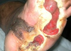
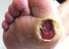
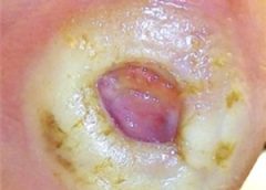
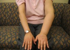
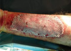
Around 6.5 million patients in the U.S. suffer from chronic wounds, such as pressure injuries or ulcers. Treatment costs $25 billion each year, representing a sizable and growing problem. Despite the wide impact of chronic wounds, it’s rare to see specialized, effective wound care delivered across the care continuum.

A chronic non-healing wound is a surrogate marker for illness. These patients require holistic management of their co-morbidities and continuity across care settings.

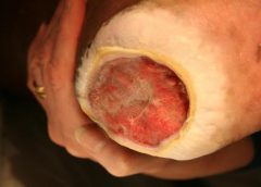
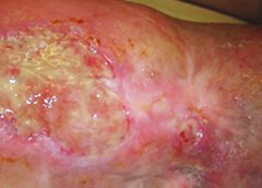
Despite this, a great deal of emphasis has been placed on treating wounds as singular events, managed topically with expensive dressings and support surfaces. This is only a small part of wound healing.

As a physician focused solely on wound care, I have learned that we must shift the focus from simply treating the wound to treating the wounded patient. The impact in the post-acute care setting in particular is worthy of evaluation and discussion, as up to 29% of patients in long-term care facilities will experience a pressure ulcer, posing serious legal, financial, and staffing implications.

For those providers working outside long-term care, there is little understanding of challenges facing LTC providers. Acute providers do not often ask, for example, how are my LTC partners reimbursed? How are they staffed? What are the requirements and regulatory pressures they face? Asking these questions would facilitate a more productive dialogue with a focus on collaborative prevention, rather than waiting until a chronic wound occurs in the LTC setting.

Creating an integrated wound care community

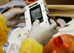
To address the needs of the present and growing population of patients with chronic wounds, Healogics developed an integrated wound care community model, to coordinate the wound healing process across all care settings. The program utilizes Healogics Specialty Physicians, a subspecialty group of physicians and providers with extensive training solely focused on wound care.

HSPs provide expert inpatient consultation and ensure safe transition of patients out of the hospital into the appropriate care setting. Because HSPs see the patient regardless of post-discharge venue, patients receive the same quality of care whether they are going home, to a skilled-nursing, assisted living, or LTC setting. Because chronic wounds are surrogate markers for illness, we have realized it’s essential to have an integrated, multi-setting, and multi-disciplinary process to treat the patient and their co-morbidities.

Data collected at a pilot IWCC site in the Midwestern U.S. from 2014 to 2016 revealed very positive trends for chronic wound patients. In the acute care setting, the average length of stay decreased from 9.41 days to 5.64 days, and total cost of care per patient was reduced from $10,670 to $7,248.

We’re excited by these promising results, which were revealed at the American College of Wound Healing and Tissue Repair Conference last December. We look forward to refining and expanding the model by helping our partners in acute and LTC settings standardize their practices, use evidence-based clinical guidelines, mobilize technologies and processes, and pay critical attention to patient safety and value-based outcomes.

When it comes to wound healing, no venue of care should operate alone—an integrated solution that creates continuity for the patient is critical. There are four things LTC facilities can do to break down the silos:

Read more at McKnight’s

Read More

NYU docs use machine learning

Lymphedema causes unsightly swelling in the arms and legs. But researchers Mei Fu and Yao Wang have an idea for catching early symptoms sooner.

Researchers at NYU’s Tandon School of Engineering have teamed up with those from the university’s Rory Meyers College of Nursing to develop a machine-learning algorithm that could help detect a lymphatic system disease before doctors are able to.

There is no cure for lymphedema, only physical exercises that can keep the symptoms in check.

Early detection of the disease would allow for physical therapy that could theoretically stop the disease’s progress enough to never allow it to develop.

“Machine learning will help us to develop an algorithm to determine a patient’s status or predict if they will have a measurable symptom later on,” explained Mei Fu, an associate professor at NYU’s Rory Meyers College of Nursing, by telephone last week. “Each time the patients enter the data, the algorithm will teach itself. Later on, machine learning will probably help us say which treatment is better for which kind of patients.”

Read more at Technically Brooklyn

Read More

One Doctor Exploring Wound Care on Earth and in Space

In laboratories all across the globe, scientists are uncovering new and exciting breakthroughs in the realm of wound healing.

For instance, a team out of Texas is blinding bacteria to prevent their spread. Meanwhile, a collective of doctors from the U.K. recently developed some intriguing new vacuum tech to treat chronic ulcers. There’s even been research into drug treatments, like how opioids may actually prevent proper wound care.

Each team has taken a different approach or tackled a unique situation or medical ailment, and that ensures a more well-rounded coverage that helps a larger pool of patients. However, few scientists have a more grand scope than Ronke Olabisi, a professor of biomedical engineering at Rutgers University.

Reaching for the stars

As the university explained in a recent press release, Olabisi is hard at work on several projects aimed at improving wound healing both on earth and during manned space missions. During space travel, especially as astronauts spend months at a time in stations, the lack of gravity has a huge impact on the human body. Muscle and bones will actually start to deteriorate, and tissues will lose much of their elasticity. Olabisi’s main goal is to study in-depth why this occurs and how to fix, and she believes she can apply much of the same knowledge to wound care on Earth.

Read more at Advanced Tissue

Read More

Accuracy of the Ankle-brachial Index in the Assessment of Arterial Perfusion of Heel Pressure Injuries

Abstract: Background. The evaluation and treatment of heel pressure injuries are a significant and expensive sequela of the aging population. Although the workup of patients with lower extremity tissue loss usually involves an assessment of the arterial blood flow by means of noninvasive vascular testing, the results may be misleading in patients with heel pressure injuries when the ankle-brachial index (ABI) does not provide direct information about perfusion of the rearfoot. The objective of this retrospective, observational investigation was to determine if noninvasive vascular testing provides accurate and reliable results in patients with heel pressure injuries. (more…)

Read More

Wound Photography – How it Benefits Clinical Documentation

wound photography

Accurate assessment and documentation of wounds is essential for developing a comprehensive plan of care. Photography now plays a key role in wound care. The use of digital photography has enhanced the reliability and accuracy of wound documentation. Though a wound assessment in patient files includes details such as location, depth, odor, condition of surrounding tissue and other details, a visual record can be worth even more.

Digital photography is becoming a more prevalent documentation tool. According to an article published in McKnight’s, forensic nursing experts recommend using photographs to document injury. The photos show both how an injury occurred and how it is healing.

The National Pressure Ulcer Advisory Panel (NPUAP) also supports photography as a more accurate means for assessment of wound dimensions and wound base over time.

A visual confirmation to the written record, these images:

  • Facilitate better diagnosis
  • Enhance clinical documentation
  • Help to monitor the progress of wound healing
  • Help prevent litigation in wound management
  • Allow inter-disciplinary communication among the wound care team

Read more at Wound Wizard

Read More

Management of Patients With Venous Leg Ulcers

It is well documented that the prevalence of venous leg ulcers (VLUs) is increasing, coinciding with an ageing population. Accurate global prevalence of VLUs is difficult to estimate due to the range of methodologies used in studies and accuracy of reporting.1 Venous ulceration is the most common type of leg ulceration and a significant clinical problem, affecting approximately 1% of the population and 3% of people over 80 years of age2 in westernised countries. Moreover, the global prevalence of VLUs is predicted to escalate dramatically, as people are living longer, often with multiple comorbidities. Recent figures on the prevalence of VLUs are based on a small number of studies, conducted in Western countries, and the evidence is weak. However, it is estimated that 93% of VLUs will heal in 12 months, and 7% remain unhealed after five years.3 Furthermore, the recurrence rate within 3 months after wound closure is as high as 70%.4-6 Thus, cost-effective adjunct evidence-based treatment strategies and services are needed to help prevent these ulcers, facilitate healing when they occur and prevent recurrence.

The impact of a VLU represents social, personal, financial and psychological costs on the individual and further economic drain on the health-care system. This brings the challenge of providing a standardised leg ulcer service which delivers evidence-based treatment for the patient and their ulcer. It is recognised there are variations in practice and barriers preventing the implementation of best practice. There are patients not receiving appropriate and timely treatment in the initial development of VLUs, effective management of their VLU and preventing recurrence once the VLU has healed.

Health-care professionals (HCPs) and organisations must have confidence in the development process of clinical practice guidelines and have ownership of these guidelines to ensure those of the highest quality guide their practice. These systematic judgments can assist in policy development, and decision making, improve communication, reduce errors and improve patient outcomes.

Read more at Journal of Wound Care

Read More

Reduction of 50% in Diabetic Foot Ulcers With Stem Cells

Diabetic Foot Ulcers

MUNICH — Local injection of mesenchymal stem cells derived from autologous bone marrow shows promise in healing recalcitrant neuropathic diabetic foot ulcers, a novel study from Egypt shows.

Presenting the results at the European Association for the Study of Diabetes (EASD) 2016 Annual Meeting, Ahmed Albehairy, MD, from Mansoura University, Egypt, said: “In patients who received the mesenchymal stem cells, ulcer reduction was found to be significantly higher compared with patients on conventional treatment after both 6 weeks and 12 weeks of follow-up. This is despite the fact that initial ulcer size was larger in the stem-cell–treated group.” (more…)

Read More

Better Skin Grafts – take only one layer

skin grafts take one layer

Research shows that a skin-graft harvesting system aids chronic wound recovery and reduces care costs by accelerating the healing process.

More than six million cases of chronic wounds cost $20 billion each year in the United States. Diabetic ulcers, pressure sores, surgical site wounds, and traumatic injuries to high-risk patients account for most wounds that won’t heal. (more…)

Read More
1 2 3 14