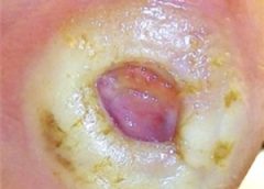
Diabetic foot ulcers stem from multiple factors, including peripheral neuropathy, high plantar pressures, decreased vascularity, and impaired wound healing. Contributing significantly to morbidity, they may cause limb loss and death. (See Foot ulcers and diabetes.)
Initially, hydrocolloid dressings were developed to function as part of the stomal flange. Based on their success in protecting peristomal skin, they were introduced gradually into other areas of wound care. They contain wafers of gel-forming polymers, such as gelatin, pectin, and cellulose agents, within a flexible water-resistant outer layer. The wafers absorb wound exudate, forming a gel and creating a moist healing environment. (more…)
Factors affecting medication adherence in patients with diabetes identified
Factors associated with better adherence to antidiabetic medications taken by patients with diabetes include older age, male sex, higher education, higher income, use of mail-order vs. retail pharmacies, primary care vs. nonendocrinology specialist prescribers, higher daily total pill burden, and lower out-of-pocket costs. (more…)
By Nancy Morgan, MBA, BSN, RN, WOC, WCC, CWCMS, DWC
Each month, Apple Bites brings you a tool you can apply in your daily practice.
Description
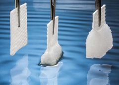
• Semipermeable polyurethane foam dressing
• Nonadherent and nonlinting
• Hydrophobic or waterproof outer layer
• Provides moist wound environment
• Permeable to water vapor but blocks entry of bacteria and contaminants
• Available in various thicknesses with or without adhesive borders
• Available in pads, sheets, and cavity dressings (more…)
BY: NANCY MORGAN, RN, BSN, MBA, WOCN, WCC, CWCMS, DWC
What exactly is wound exudate? Also known as drainage, exudate is a liquid produced by the body in response to tissue damage. We want our patients’ wounds to be moist, but not overly moist. The type of drainage can tell us what’s going on in a wound.
Let’s look at the types of exudates commonly seen with wounds. (more…)
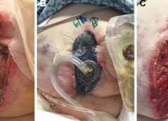
Negative pressure wound therapy (NPWT) uses negative pressure to draw wound edges together, remove edema and infectious material, and promote perfusion and granulation tissue development. The tissue stretch and compression created by negative pressure during NPWT promotes tissue perfusion and granulation tissue development through angiogenesis, cellular proliferation, fibroblast migration, increased production of wound healing proteins, and reduction of wound area. NPWT has been used to improve healing in a variety of wounds, including traumatic injuries, surgical wounds, pressure ulcers, diabetic foot ulcers, and venous stasis ulcers. (more…)
According to the National Cancer Institute, an estimated 1.6 million new cases of cancer will have been diagnosed in the United States in 2015. During the course of their disease, most cancer patients receive radiation therapy.
Delivering high energy in the form of waves or particles, radiation therapy alters the DNA of cancer cells, causing their death. Radiation can be administered either externally or internally (through materials placed into the body). It’s given in fraction doses, with the total recommended dose divided into daily amounts. Treatment, including the total dose, is determined on an individual basis.
Although improvements have been made in delivery of radiation therapy, approximately 95% of patients who receive it experience a skin reaction. What’s more, radiation therapy commonly is given concurrently with chemotherapy or targeted therapy to improve survival, which increases the toxicity risk. (more…)
The authors of the article explain evidence-based practice and provide useful definitions for key terms. They then provide a list of eight questions to use when evaluating SRs and practical tips such as how to search for SR and MA studies. The article finishes with a list of eight interventions supported by the most evidence: hydrocolloidal dressings, honey, biosynthetic dressings, iodine complexes, silver compounds, hydrogels, foam dressings, and negative pressure wound therapy. (more…)
Critical limb ischemia may not increase mortality risk in patients with diabetes
Diabetic patients with critical limb ischemia (CLI) who are assessed quickly and treated aggressively do not have an increased risk of long-term cardiac mortality, according to a study in Diabetes Care.(more…)
In health care, we frequently use the terms formulary and protocol interchangeably even though they have different meanings. A formulary is an official list of available dressings, products, and medications. A protocol is a roadmap or guideline on how to use the formulary.
Formularies became popular several years ago when reimbursement changed to bundling and wound-product costs were included in the routine cost of care rather than separately billable. In an effort to control costs, hospitals, home health agencies, and long-term care facilities began exclusive partner agreements with supply and buying groups. (“You use our products exclusively and we’ll give you a huge discount on cost.”)
A good formulary not only can help save money. It can also assist in streamlining care delivery, reducing waste, and directing treatment decisions. But on the flip side, using formularies can have disastrous results. I realized this last week while speaking on the phone with a wound clinician who’d called to ask for wound treatment ideas for a hospice patient. As she described the situation, it became apparent that the patient’s symptoms definitely pointed to high levels of bacteria in the wound. As I began sharing recommendations for treatment ideas, she kept responding: “Nope. Can’t use that, not on our formulary.” “Nope, not on formulary.” The only options available on her hospice formulary were hydrocolloid, hydrogel, or foam dressings, none of which had antibacterial properties.
Providing an appropriate standard of care shouldn’t be dictated by a formulary, and choosing substandard care just because the patient is in hospice isn’t acceptable or appropriate. Evidence-based guidelines, wound characteristics, underlying complications, and patient care goals should dictate management and treatment.
To ensure your formulary is adequate, determine if it includes a variety of product categories, and negotiate the ability to go off formulary if needed. Although cost control is essential, clinicians need access to products and therapies that yield positive outcomes. One size doesn’t fit all in wound care.
Donna Sardina, RN, MHA, WCC, CWCMS, DWC, OMS
Editor-in-Chief Wound Care Advisor
Cofounder, Wound Care Education Institute
Plainfield, Illinois
By Nancy Chatham, MSN, RN, ANP-BC, CWOCN, CWS, and Carrie Carls, BSN, RN, CWOCN, CHRN
Moisture-related skin breakdown has been called many things-perineal dermatitis, irritant dermatitis, contact dermatitis, heat rash, and anything else caregivers could think of to describe the damage occurring when moisture from urine or stool is left on the skin. At a 2005 consensus conference, attendees chose the term incontinence-associated dermatitis (IAD).
IAD can be painful, hard to properly identify, complicated to treat, and costly. It’s part of a larger group of moisture-associated skin damage that also includes intertrigo and periwound maceration. IAD prevalence and incidence vary widely with the care setting and study design. Appropriate diagnosis, prompt treatment, and management of the irritant source are crucial to long-term treatment.
Causes
IAD stems from the effects of urine, stool, and containment devices on the skin. The skin’s pH contributes to its barrier functions and defenses against bacteria and fungus; ideal pH is 5.0 to 5.9. Urine pH ranges from 4.5 to 8.0; the higher range is alkaline and contributes to skin damage.
Skin moisture isn’t necessarily damaging. But when moisture that contains irritating substances, such as alkaline urine, contacts the skin for a prolonged period, damage can occur. Urine on the skin alters the normal skin flora and increases permeability of the stratum corneum, weakening the skin and making it more susceptible to friction and erosion. Fecal incontinence leads to active fecal enzymes on the skin, which contribute to skin damage. Fecal bacteria can penetrate the skin, increasing the risk of secondary infection. Wet skin has a lower temperature than dry skin; wet skin under a pressure load has less blood flow than dry skin.
Containment devices, otherwise known as adult diapers or briefs, are multilayer disposable garments containing a superabsorbent polymer. The polymer is designed to wick and trap moisture in the containment device. This ultimately affects the skin by trapping heat and moisture, which may cause redness and inflammation that can progress to skin erosion. This trapping can lead to increased pressure against the skin, especially if the device has absorbed liquid and remains in contact with the skin.
Categorizing IAD
IAD is categorized as mild, moderate, or severe. (See Picturing IAD by clicking the PDF icon above.)
Screening for IAD
Screen the patient’s skin for persistent redness, inflammation, rash, pain, and itching at least daily. To differentiate IAD from pressure ulcers, keep in mind that:
IAD can occur wherever urine or stool contacts the skin. In contrast, pressure ulcers arise over bony prominences in the absence of moisture.
With IAD, affected skin is red or bright red. With a pressure ulcer, skin may take on a bluish purple, red, yellow, or black discoloration.
The skin-damage pattern in IAD usually is diffuse. With a pressure ulcer, edges are well defined.
The depth of IAD-related skin damage usually is partial-thickness without necrotic tissue. With a pressure ulcer, skin damage depth may vary.
Preventing IAD
The three essentials of IAD prevention are to cleanse, moisturize, and protect.
Cleanse the skin with a mild soap that’s balanced to skin pH and contains surfactants that lift stool and urine from the skin. Clean the skin routinely and at the time of soiling. Use warm (not hot) water, and avoid excess force and friction to avoid further skin damage.
Moisturize the skin daily and as needed. Moisturizers may be applied alone or
incorporated into a cleanser. Typically, they contain an emollient such as lanolin to replace lost lipids in the stratum corneum.
To protect the skin, apply a moisture-barrier cream or spray if the patent has significant urinary or fecal incontinence (or both). The barrier may be zinc-based, petrolatum-based, dimethicone-based, an acrylic polymer, or another type. Consider using an algorithm developed by wound and skin care specialists that’s customized for skin care products your facility uses. (See Skin care algorithm by clicking the PDF icon above.)
If the treatment protocol fails, the patient should be referred to an appropriate skin care specialist promptly.
To help prevent urine or stool from contacting the patient’s skin, consider using a male external catheter, a female urinary pouch, a fecal pouch, or a bowel management system. Avoid containment devices. If the patient has a containment pad, make sure it’s highly absorbent and not layered, to decrease pressure under the patient.
Managing IAD
A comprehensive multidisciplinary approach to IAD is essential to the success of any skin care protocol. Identify skin care champions within your facility and educate them on IAD. Incorporating administrators, physicians, nursing staff, therapists, and care assistants makes implementation of protocols and algorithms within an institution seamless.
Administrators support the skin care program in the facility, including authorizing a budget so product purchases can be made. The certified wound clinician is the team expert regarding skin care, incontinence, prevention, and product recommendation. The physician oversees protocol development and evaluates and prescribes additional treatment when a patients fails to respond to treatment algorithms. Nursing staff identify patients at risk, incorporate the algorithm into the patient’s plan of care, and direct care
assistants. Therapists address function, strength, and endurance issues to improve the patient’s self-care abilities in activities of daily living to manage or prevent episodes of incontinence.
In severe inflammation, topical dressings, such as alginates and foam dressings, may be used along with topical corticosteroids. In complex IAD, antifungals or antibiotics may be required if a secondary fungal or bacterial infection is suspected.
Additional diagnostic tests may be done to identify and treat secondary infections. These tests may include skin scraping, potassium hydroxide test or Gram’s stain for fungal components, or a swab culture and sensitivity for bacterial infections. If your patient has a suspected secondary fungal or bacterial infection, use appropriate treatments for the full course of recommended therapy. In severe secondary fungal infection, an oral agent may be added to topical therapy. If cost is a concern, consider using a pharmacy knowledgeable about compounding for topical combination therapies.
Referrals and education
For assessment and treatment of under-lying incontinence, refer the patient to a continence specialist if appropriate. Teach the patient strategies for managing incontinence through dietary measures, toileting programs, pelvic-floor muscle training, clothing modification, and mobility aids.
Selected references
Beguin A, Malaquin-Pavan E, Guihaire C, et al., Improving diaper design to address incontinence associated dermatitis. BMC Geriatrics. 2010;10:86. http://www.biomedcentral.com/1471-2318/10/86. Accessed March 15, 2012.
Black JM, Gray M, Bliss DZ, et al. MASD part 2: incontinence-associated dermatitis and intertriginous dermatitis. J Wound Ostomy Continence Nurs. 2011; 38(4):359-370.
Bliss DZ, Zehrer C, Savik K, et al. An economic evaluation of four skin damage prevention regimens in nursing home residents with incontinence: economics of skin damage prevention. J Wound Ostomy Continence Nurs. 2007;34(2):143-152.
Denat Y, Khorshid L. The effect of 2 different care products on incontinence-associated dermatitis in patients with fecal incontinence. J Wound Ostomy Continence Nurs. 2011;38(2):171-176.
Doughty DB. Urinary and Fecal Incontinence: Current Management Concepts. 3rd ed. St. Louis, MO: Mosby Elsevier; 2006.
Gray, M. Optimal management of incontinence-associated dermatitis in the elderly. Am J Clin Dermatol. 2010;11(3):201-210.
Gray M, Beeckman D, Bliss DZ, et al. Incontinence-associated dermatitis: a comprehensive review and update. J Wound Ostomy Continence Nurs. 2012;39(1):61-74
Gray M, Bliss DZ, Doughty DB, et al. Incontinence-associated dermatitis: a consensus. J Wound Ostomy Continence Nurs. 2007;34(1):45-54.
Gray M, Bohacek L, Weir D, et al. Moisture vs pressure: making sense out of perineal wounds. J Wound Ostomy Continence Nurs. 2007;34(2):134-42.
Junkin J, Lerner-Selekof JL. Prevalence of incontinence and associated skin injury in the acute care inpatient. J Wound Ostomy Continence Nurs. 2007;34(3):260-269.
Landefeld CS, Bowers BJ, Feld AD, et al. National Institutes of Health state-of-the-science conference statement: prevention of fecal and urinary incontinence in adults. Ann Intern Med. 2008;148(6):449-458.
Langemo D, Hanson D, Hunter S, et al. Incontinence and incontinence-associated dermatitis. Adv Skin Wound Care. 2011;24(3):126-142.
Nancy Chatham is an advanced practice nurse at Passavant Physician Associates in Jacksonville, Illinois. Carrie Carls is the nursing director of advanced wound healing and hyperbaric medicine at Passavant Area Hospital in Jacksonville, Illinois.
A new study shows a clear association between the prophylactic use of five-layer foam sacral dressings and reductions in pressure injury rates. Specifically, the study looked at the prophylactic use of Mölnlycke’s Mepilex® Border Sacrum dressing in the acute care setting over a six-year period (2010-2015). (more…)
The cutting-edge of wound care is a progressively flexible one, where textiles, foams, and films are applied to wound management technology with the goal of synergistic physiological function. These innately intuitive materials underpin the emerging medical solutions that practitioners and their patients are finding more effective than traditional wound care and closure methods. With an aging population more frequently seeking medical care and a surge in diabetes diagnoses, market analysts predict a continuing rise in demand for advanced wound care management products, fueling an annual industry growth rate of 6.4% over the next five years. (more…)