As wound care clinicians, we know that an interdisciplinary, holistic approach to prevention and management of a wound is crucial to positive outcomes, no matter where the patient is being seen. Yet too often when a patient transfers from one care setting to another, the only wound information that’s communicated is the current topical treatment. Most transfer forms only include generic spaces for “any skin concerns” and “treatments,” with no prompts for obtaining additional information. In fact, clinicians in many care settings frequently report they had no idea the patient had a wound until he or she was admitted.
Here’s how you can get the information you need to best care for the patient being transferred. (more…)
By Beth Hoffmire Heideman, MSN, BSN, RN, WCC, DWC, OMS
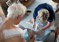
Fifty shades of wound care at home refers to treating the whole patient and the patient’s caregiving supporters—not just the wound. Only by understanding the nuances, or shades, of a patient and his or her environment can clinicians best achieve desired outcomes.
Wound healing in home care depends on teamwork. Members of the team must understand the unique situation of delivering care in the home and how to help patients adhere to the plan of care. (more…)
A pressure ulcer that a patient acquires in your facility or a patient’s existing pressure ulcer that worsens puts your organization at risk for regulatory citations as well as litigation. Unless you can prove the pressure ulcer was unavoidable, you could find yourself burdened with citations or fines, or could even end up in court. (more…)
A pressure ulcer that a patient acquires in your facility or a patient’s existing pressure ulcer that worsens puts your organization at risk for regulatory citations as well as litigation. Unless you can prove the pressure ulcer was unavoidable, you could find yourself burdened with citations or fines, or could even end up in court.
In 2010, the National Pressure Ulcer Advisory Panel (NPUAP) hosted a multidisciplinary conference to establish a consensus on whether all pressure ulcers are avoidable.
Hospital pressure-ulcer comparison data not accurate Performance scores for rates of hospital-acquired pressure ulcers might not be appropriate for comparing hospitals, according to a study in the Annals of Internal Medicine. “Hospital report cards for hospital-acquired pressure ulcers: How good are the grades?,” funded by the Agency for Healthcare Research and Quality, analyzed 2 million all-payer administrative records from 448…
A variety of resources to end the year and take you into 2014. On the road again Give your patients with an ostomy this information from the Transportation Security Administration to help them navigate airport screening: • You can be screened without having to empty or expose your ostomy, but you need to let the officer conducting the screening know…
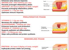
By Nancy Collins, PhD, RD, LD/N, FAPWCA, and Allison Schnitzer Nutrition is a critical factor in the wound healing process, with adequate protein intake essential to the successful healing of a wound. Patients with both chronic and acute wounds, such as postsurgical wounds or pressure ulcers, require an increased amount of protein to ensure complete and timely healing of their…
By Jeri Lundgren, BSN, RN, PHN, CWS, CWCN A pressure ulcer that a patient acquires in your facility or a patient’s existing pressure ulcer that worsens puts your organization at risk for regulatory citations as well as litigation. Unless you can prove the pressure ulcer was unavoidable, you could find yourself burdened with citations or fines, or could even end…
By Kathleen D. Pagana, PhD, RN Are you making connections that benefit your career? Are you comfortable starting a conversation at a networking session? Do you know how to exit a conversation gracefully when it’s time to move on? These are questions and concerns many clinicians share. Career success takes more than clinical expertise, management savvy, and leadership skills. Networking…
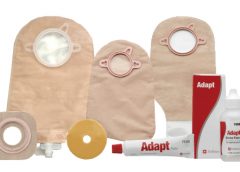
By Connie Johnson, BSN, RN, WCC, LLE, OMS, DAPWCA No matter where you work or who your distributors are, ensuring the patient has sufficient ostomy supplies can be a challenge. Whether you’re the nurse, the physician, the patient, or the family, not having supplies for treatments can heighten frustration with an already challenging situation, such as a new ostomy. Here’s…
by Donna Sardina, RN, MHA, WCC, CWCMS, DWC, OMS With uncertainty over how the Affordable Care Act (ACA) ultimately will affect operations, hospitals and other healthcare facilities are tightening up. In many areas, they’re laying off staff. In May, the healthcare industry lost 9,000 jobs—the worst month for the industry in a decade—and another 4,000 jobs were lost in July.…
Dermatologic difficulties: Skin problems in patients with chronic venous insufficiency and phlebolymphedema By Nancy Chatham, RN, MSN, ANP-BC, CWOCN, CWS; Lori Thomas, MS, OTR/L, CLT-LANA; and Michael Molyneaux, MD Skin problems associated with chronic venous insufficiency (CVI) and phlebolymphedema are common and often difficult to treat. The CVI cycle of skin and soft tissue injury from chronic disease processes can…
By Robyn Bjork, MPT, WCC, CWS, CLT-LANA Margery Smith, age 82, arrives at your wound clinic for treatment of a shallow, painful ulcer on the lateral aspect of her right lower leg. On examination, you notice weeping and redness of both lower legs, 3+ pitting edema, several blisters, and considerable denudement of the periwound skin. She is wearing tennis shoes…
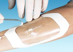
By Nancy Morgan, RN, BSN, MBA, WOC, WCC, DWC, OMS Each issue, Apple Bites brings you a tool you can apply in your daily practice. Description Hydrated polymer (hydrogel) dressings, originally developed in the 1950s, contain 90% water in a gel base, which helps regulate fluid exchange from the wound surface. Hydrogel dressing are usually clear or translucent and vary…
By Nancy Collins, PhD, RD, LD/N, FAPWCA, and Allison Schnitzer
Nutrition is a critical factor in the wound healing process, with adequate protein intake essential to the successful healing of a wound. Patients with both chronic and acute wounds, such as postsurgical wounds or pressure ulcers, require an increased amount of protein to ensure complete and timely healing of their wounds.
Elderly patients with wounds pose a special challenge because of their decreased lean body mass and the likelihood of chronic illnesses and insufficient dietary protein intake. To promote a full recovery, wound care clinicians must address the increased protein needs of wound patients, especially elderly patients. (more…)
By Tamera L. Brown, MS, RN, ACNS-BC, CWON, and Jessica Kitterman, BSN, RN, CWOCN
Pressure ulcers take a hefty toll in both human and economic terms. They can lengthen patient stays, cause pain and suffering, and increase care costs. The average estimated cost of treating a pressure ulcer is $50,000; this amount may include specialty beds, wound care supplies, nutritional support, and increased staff time to care for wounds. What’s more, national patient safety organizations and insurance payers have deemed pressure ulcers avoidable medical errors and no longer reimburse the cost of caring for pressure ulcers that develop during hospitalization. (more…)
Achieving excellent wound care outcomes can be challenging, given the growing number of high-risk patients admitted to healthcare facilities today. Many of these patients have comorbidities, such as obesity, diabetes, renal disease, smoking, chronic obstructive pulmonary disease, and poor nutritional status. These conditions reduce wound-healing ability. (more…)
The first 24 hours after a patient’s admission are critical in preventing pressure ulcer development or preventing an existing ulcer from worsening. A skin inspection, risk assessment, and temporary care plan should all be implemented during this time frame. Essentially, it’s the burden of the care setting to prove to insurers, regulators, and attorneys the pressure ulcer was present on admission and interventions were put into place to avoid worsening of the condition. Of course, patients also benefit from having their condition identified and treated promptly. (more…)
Pressure ulcers take a hefty toll in both human and economic terms. They can lengthen patient stays, cause pain and suffering, and increase care costs. The average estimated cost of treating a pressure ulcer is $50,000; this amount may include specialty beds, wound care supplies, nutritional support, and increased staff time to care for wounds. What’s more, national patient safety organizations and insurance payers have deemed pressure ulcers avoidable medical errors and no longer reimburse the cost of caring for pressure ulcers that develop during hospitalization.
Frequent debridement improves wound healing A study in JAMA Dermatology reports that frequent debridements speed wound healing. “The more frequent the debridement, the better the healing outcome,” concludes “Frequency of debridements and time to heal: A retrospective cohort study of 312 744 wounds.” The median number of debridements was two. Most of the wounds in the 154,644 patients were diabetic foot…
Here are some resources of value to your practice. National Guideline Clearinghouse The National Guideline Clearinghouse, supported by the Agency for Healthcare Research and Quality, summarizes many guidelines of interest to wound care, ostomy, and lymphedema clinicians. Here are some examples: Guideline for management of wounds in patients with lower-extremity neuropathic disease Pressure ulcer prevention and treatment protocol Lower limb…
By Gail Hebert, RN, MS, CWCN, WCC, DWC, LNHA, OMS; and Rosalyn Jordan, BSN, RN, MSc, CWOCN, WCC, OMS Imagine your physician has just told you that your rectal pain and bleeding are caused by invasive colon cancer and you need prompt surgery. She then informs you that surgery will reroute your feces to an opening on your abdominal wall.…
By Rose O. Sherman, EdD, RN, NEA-BC, FAAN Unfortunately, most clinicians can’t avoid having to work with difficult people. However we can learn how to be more effective in these situations, keeping in mind that learning to work with difficult people is both an art and a science. How difficult people differ from the rest of us We can all…
By Tamera L. Brown, MS, RN, ACNS-BC, CWON, and Jessica Kitterman, BSN, RN, CWOCN Pressure ulcers take a hefty toll in both human and economic terms. They can lengthen patient stays, cause pain and suffering, and increase care costs. The average estimated cost of treating a pressure ulcer is $50,000; this amount may include specialty beds, wound care supplies, nutritional…
By Gregory S. Kopp, RN, MN, MHA A new job can be stimulating, but it can also be stressful. Not only will you have new responsibilities, but you’ll also have a new setting, new leaders, and new colleagues. And the quicker you can figure out who’s who and what’s what—without stepping on anyone’s toes—the better off you’ll be. But establishing…
By Ronnel Alumia, BSN, RN, WCC, CWCN, OMS Achieving excellent wound care outcomes can be challenging, given the growing number of high-risk patients admitted to healthcare facilities today. Many of these patients have comorbidities, such as obesity, diabetes, renal disease, smoking, chronic obstructive pulmonary disease, and poor nutritional status. These conditions reduce wound-healing ability.
By Janice M. Beitz, PhD, RN, CS, CNOR, CWOCN, CRNP Quality patient education is essential for comprehensive health care and will become reimbursable under healthcare reform in 2014. However, it’s difficult to provide effective education when time for patient interactions is limited. You can enhance your instruction time—and make your teaching more memorable—by using the techniques of analogy and metaphor.
By Jeri Lundgren, BSN, RN, PHN, CWS, CWCN The first 24 hours after a patient’s admission are critical in preventing pressure ulcer development or preventing an existing ulcer from worsening. A skin inspection, risk assessment, and temporary care plan should all be implemented during this time frame. Essentially, it’s the burden of the care setting to prove to insurers, regulators,…
By: Donna Sardina, RN, MHA, WCC, CWCMS, DWC, OMS Why is it that the people who are the most caring toward others neglect their own needs? Have you noticed this? I’ve seen it time and time again. The healthcare worker who’s always the last to leave work, who always volunteers to work those extra shifts so patient care won’t be…
By Rosalyn S. Jordan, RN, BSN, MSc, CWOCN, WCC, OMS; and Judith LaDonna Burns, LPN, WCC, DFC
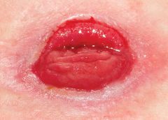
About 1 million people in the United States have either temporary or permanent stomas. A stoma is created surgically to divert fecal material or urine in patients with GI or urinary tract diseases or disorders.
A stoma has no sensory nerve endings and is insensitive to pain. Yet several complications can affect it, making accurate assessment crucial. These complications may occur during the immediate postoperative period, within 30 days after surgery, or later. Lifelong assessment by a healthcare provider with knowledge of ostomy surgeries and complications is important. (more…)
One of the worst fears of a wound care clinician is inadvertently compressing a leg with critical limb ischemia—a condition marked by barely enough blood flow to sustain tissue life. Compression (as well as infection or injury) could lead to necrosis, the need for amputation, or even death. The gold standard of practice is to obtain an ankle-brachial index (ABI) before applying compression. However, recent research and expert opinion indicate an elevated or normal ABI is deceptive in patients with advanced diabetes. What’s worse, in the diabetic foot, skin may die from chronic capillary ischemia even when total blood perfusion is normal. For information on how to perform an ABI and interpret results, click on this link. (more…)
Most of us have had days when we jump from meeting to meeting and at the end of the day wonder, “Did I get anything accomplished or am I more behind than ever?”
Many clinicians tell me that although their wound team meets regularly, the meetings aren’t meaningful enough, leaving the team still facing issues with their wound care program. As a consultant, when I review the wound team agenda, it’s typically missing one or more of four key ingredients:
appropriate member representation
proactive approach that highlights prevention
review of the plan of care and update of the medical record
review of supplies and products. Here’s a closer look at each of these ingredients.
Build a top team
Having the appropriate members on the wound care team is the first ingredient for success. A comprehensive, interdisciplinary team approach is the key to preventing skin breakdown and ensuring good clinical outcomes for residents with skin breakdown. Teams should include representation from nursing, dietary, and physical and occupational therapy, as well as a nurse practitioner or physician.
Nursing representation should include nurses from all three shifts and nursing assistants, who are too often missing from the team. Keep in mind that when it comes to preventing pressure ulcers, nursing assistants carry out most of interventions (for example, turning, incontinence management, heel lift). Even when a patient has a wound, the only intervention carried out by the nurses is the topical treatment; nursing assistants perform all other interventions necessary to ensure healing. Clinicians who empower nursing assistants to have a strong influence with the wound care team—and the program—tend to have very successful prevention programs and good clinical outcomes.
Think prevention
The second key ingredient is prevention. Most wound team meetings only discuss the patients with wounds, missing the bigger goal of preventing wounds in the first place. Once the patients with wounds are discussed, the team should review all high-risk patients to ensure proper preventative measures are in place and care planned. All patients should be quickly reviewed for evidence of:
decline or change in mobility and activity
new onset or change in continence status
decline in nutritional status
decline or change in cognition.
Any triggers in these areas should prompt a review of the plan of care to ensure they are being effectively addressed.
Review and update the plan
The third key ingredient for success is to use meeting time to review and update the plan of care. I’ve observed highly productive meetings and great discussions of the care the facility is providing. Then I review the medical record and discover that none of the interventions discussed are on the plan of care. Always review the patient’s plan of care to ensure it’s accurate, reflects all interventions, and is up to date. This will give you peace of mind that the medical record reflects all the good work you’re doing and helps make the team meetings feel productive.
Discuss products and supplies
The fourth key ingredient is to take the time to quickly discuss current wound care supplies and products with the team. Ask the team if the current supplies are user-friendly, are adequate, provide good outcomes, and are in good working condition.
Many times staff will not say how they’re struggling with, modifying, or not using something until they’re asked. Remember that the most expensive product is the one that doesn’t work or doesn’t get used.
A recipe for success
Using these four key ingredients will lead you to a successful wound team meeting—and a successful program. The mix may not solve your too-many-meetings days, but will give you peace of mind that at least one meeting is productive.
Jeri Lundgren is director of clinical services at Pathway Health in Minnesota. She has been specializing in wound prevention and management since 1990.