By Rosalyn S. Jordan, RN, BSN, MSc, CWOCN, WCC, OMS; and Judith LaDonna Burns, LPN, WCC, DFC
About 1 million people in the United States have either temporary or permanent stomas. A stoma is created surgically to divert fecal material or urine in patients with GI or urinary tract diseases or disorders.
A stoma has no sensory nerve endings and is insensitive to pain. Yet several complications can affect it, making accurate assessment crucial. These complications may occur during the immediate postoperative period, within 30 days after surgery, or later. Lifelong assessment by a healthcare provider with knowledge of ostomy surgeries and complications is important.
Immediately after surgery, a healthy GI stoma appears red, moist, and shiny. Edema of the stoma is expected for the first 6 to 8 weeks. A healthy urinary stoma is pale or pink, edematous, moist, and shiny. Usually, it shrinks to about one-third the initial size after the first 6 to 8 weeks as edema subsides. The stoma warrants close observation as pouching types and sizes may need to be changed during this time. Teach the patient and family caregivers to report changes or signs and symptoms of stoma complications to a healthcare provider. If complications are recognized early, the problem may be resolved without surgical intervention.
Stoma complications range from a simple, unsightly protrusion to conditions that require emergency treatment and possible surgery. Clinicians must be able to recognize complications and provide necessary treatment and therapy early. Complications include parastomal hernias, stoma trauma, mucocutaneous separation, necrosis, prolapse, retraction, and stenosis. Although one complication can lead to and even promote others, all require attention and treatment.
Parastomal hernia
A parastomal hernia involves an ostomy in the area where the stoma exits the abdominal cavity. The intestine or bowel extends beyond the abdominal cavity or abdominal muscles; the area around the stoma appears as a swelling or protuberance. Parastomal hernias are incisional hernias in the area of the abdominal musculature that was incised to bring the intestine through the abdominal wall to form the stoma. They may completely surround the stoma (called circumferential hernias) or may invade only part of the stoma.
Parastomal hernias can occur any time after the surgical procedure but usually happen within the first 2 years. Recurrences are common if the hernia needs to be repaired surgically. Risk factors may be patient related or technical. Patient-related risk factors include obesity, poor nutritional status at the time of surgery, presurgical steroid therapy, wound sepsis, and chronic cough. Risk factors related to technical issues include size of the surgical opening and whether surgery was done on an emergency or elective basis.
Parastomal hernias occur in four types. (See Types of parastomal hernias by clicking the PDF icon above.) Initially, a parastomal hernia begins as an unsightly distention in the area surrounding the stoma; the hernia enlarges, causing pain, discomfort, and pouching problems resulting in peristomal skin complications that require frequent assessment. Conservative therapy is the usual initial treatment. Adjustments to the pouching system typically are required so changes in the shape of the pouching surface can be accommodated. Also, a hernia support binder or pouch support belt may be helpful. Avoid convex pouching systems; if this isn’t possible, use these systems with extreme caution. If the patient irrigates the colostomy, an ostomy management specialist should advise the patient to discontinue irrigation until the parastomal hernia resolves.
Stoma trauma
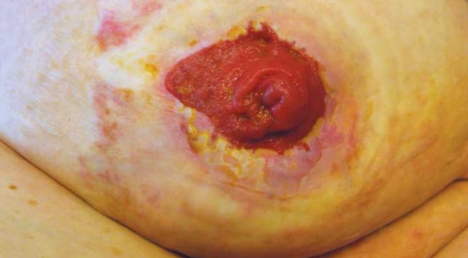
Stoma trauma occurs when the stoma is injured, typically from a laceration. Lacerations usually result from the pouch appliance or clothing. Belt-line stomas are easily traumatized and injury may occur from both clothing belts and pouch support belts. Stoma lacerations commonly result from a small opening in the flange or a misaligned pouch opening. Other causes include parastomal or stomal prolapse with possible stoma enlargement or edema.
Signs and symptoms of stoma trauma include bright red bleeding, a visible cut, and a yellowish-white linear discoloration. Lacerations may heal spontaneously. If the culprit is the pouching system, make sure nothing within the system comes in contact with the stoma. Usually direct pressure controls bleeding, but if bleeding continues, refer the patient to a physician for treatment.
Mucocutaneous separation
Mucocutaneous separation occurs when the stoma separates from the skin at the junction between the skin and the intestine used to form the stoma. Causes are related to poor wound-healing capacity, such as malnutrition, steroid therapy, diabetes, infection, or radiation of the abdominal area. Tension or tautness of the suture line also can cause mucocutaneous separation.
This complication usually arises early and can lead to other serious conditions, such as infection, peritonitis, and stomal stenosis. The area of the separation may completely surround the stoma (known as a circumferential separation), or the separation may affect only certain areas of the stoma/skin junction. The separation may be superficial or deep.
The first sign of mucocutaneous separation may be induration. Treat the separation as a wound, and apply wound-healing principles: Absorb drainage, reduce dead space, use the proper dressing, and promote wound healing. The proper dressing depends on wound depth and amount of wound drainage. Be sure to assess the wound, using the “clock method” to describe location; measure the wound area in centimeters; and describe the type of tissue in the wound bed. Be aware that slough may be present.
Treatment of the wound dictates how often the pouch is changed. A two-piece pouching system commonly is used to reduce the number of pouch changes. Cover the wound dressing with the pouching system unless the wound is infected. If infection is present, let the wound drain into the pouch and heal by secondary intention. Don’t use a convex pouching system, because this may cause additional injury to the mucocutaneous junction.
Stoma necrosis
Blood flow and tissue perfusion are essential to stoma health. Deficient blood flow causes stoma necrosis. A stoma may be affected by both arterial and venous blood compromise. The cause of necrosis usually relates to the surgical procedure, such as tension or too much trimming of the mesentery, or the vascular system that provides blood flow to the intestine. Other causes of vascular compromise include hypovolemia, embolus, and excessive edema.
Stoma necrosis usually occurs within the first 5 postoperative days. The stoma appears discolored rather than red, moist, and shiny. Discoloration may be cyanotic, black, dark red, dusky bluish purple, or brown. The stoma mucosa may be hard and dry or flaccid. Also, the stoma has a foul odor. Associated complications may include stoma retraction, mucocutaneous separation, stoma stenosis, and peritonitis.
Report signs and symptoms to the primary care provider immediately. Superficial necrosis may resolve with necrotic tissue simply sloughing away. But if tissue below the fascial level is involved, surgery is necessary. A transparent two-piece pouching system is recommended for frequent stoma assessment. The pouch may need to be resized often.
Stoma prolapse
A stoma prolapse occurs when the stoma moves or becomes displaced from its proper position. The proximal segment of the bowel intussuscepts and slides through the orifice of the stoma, appearing to telescope. This occurs more often in loop transverse colostomies. A prolapsed stoma increases in both length and size. Prolapse may be associated with stoma retraction and parastomal hernias.
Causes of stoma prolapse include large abdominal-wall openings, inadequate bowel fixation to the abdominal wall during surgery, increased abdominal pressure, lack of fascial support, obesity, pregnancy, and poor muscle tone.
Unless the patient complains of pain, has a circulatory problem, or has signs or symptoms of bowel obstruction, conservative treatment is used for uncomplicated stoma prolapse. The prolapse usually can be reduced with the patient in a supine position. After reduction, applying a hernia support binder often helps. Also, a stoma shield can be used to protect the stoma. A prolapsed stoma may require a larger pouch to accommodate the larger stoma. Some clinicians use cold compresses and sprinkle table sugar on the stoma; the sugar provides osmotic therapy or causes a fluid shift across the stoma mucosa and reduces edema.
Stoma retraction
The best-formed stoma protrudes about 2.5 cm, with the lumen located at the top center or apex of the stoma to guide the effluent flow directly into the pouch. In stoma retraction, the stoma has receded about 0.5 cm below the skin surface. Retraction may be circumferential or may occur in only one section of the stoma.
The usual causes of stoma retraction are tension of the intestine or obesity. Stoma retraction during the immediate postoperative period relates to poor blood flow, obesity, poor nutritional status, stenosis, early removal of a supporting device with loop stomas, stoma placement in a deep skinfold, or thick abdominal walls. Late complications usually result from weight gain or adhesions. Stoma retraction is most common in patients with ileostomies.
A retracted stoma has a concave, bowl-shaped appearance. Retraction causes a poor pouching surface, leading to frequent peristomal skin complications. Typical therapy is use of a convex pouching system and a stoma belt. If obtaining a pouch seal is a problem and the patient has recurrent peristomal skin problems from leakage, stoma revision should be considered.
Stoma stenosis
Stoma stenosis is narrowing or constriction of the stoma or its lumen. This condition may occur at the skin or fascial level of the stoma. Causes include hyperplasia, adhesions, sepsis, radiation of the intestine before stoma surgery, local inflammation, hyperkeratosis, and surgical technique.
Stoma stenosis frequently is associated with Crohn’s disease. You may notice a reduction or other change in effluent output with both urinary and GI ostomies. With GI stoma stenosis, bowel obstruction frequently occurs; signs and symptoms are abdominal cramps, diarrhea, increased flatus, explosive stool, and narrow-caliber stool. The initial sign is increased flatus. With urinary stoma stenosis, signs and symptoms include
decreased urinary output, flank pain, high residual urine in conduit, forceful urine output, and recurrent urinary tract infections.
Partial or complete bowel obstruction and stoma stenosis at the fascial level
require surgical intervention. Conservative therapy includes a low-residue diet, increased fluid intake, and correct use of stool softeners or laxatives for colostomies.
Most stoma complications are preventable and result from poor stoma placement. Up to 20% of patients with stoma complications require surgical revision of the stoma. All patients with ostomies require ongoing, accurate assessment and, if needed, early intervention by trained clinicians.
Selected references
Al-Niaimi F, Lyon CC. Primary adenocarcinoma in peristomal skin: a case study. Ostomy Wound Manage. 2010;56(1):45-7.
Appleby SL. Role of the wound ostomy continence nurse in the home care setting: a patient case study. Home Healthc Nurse. 2011:29(3);169-77.
Black P. Managing physical postoperative stoma complications. Br J Nurs. 2009:18(17):S4-10.
Burch J. Management of stoma complications. Nurs Times. 2011;107(45):17-8, 20.
Butler DL. Early postoperative complications following ostomy surgery: a review. J Wound Ostomy Continence Nurs. 2009:36(5):513-9.
Husain SG, Cataldo TE. Late stomal complications. Clin Colon Rectal Surg. 2008:21(1):31-40.
Jones T, Springfield T, Brudwick M, Ladd A. Fecal ostomies: practical management for the home health clinician. Home Healthc Nurse. 2011;29(5):306-17.
Kann BR. Early stomal complications. Clin Colon Rectal Surg. 2008:21(1):23-30.
Nybaek H, Jemec GB. Skin problems in stoma patients. J Eur Acad Dermatol Venereol. 2010;24(3):249-57.
Shabbir J, Britton DC. Stomal complications: a literature overview. Colorectal Dis. 2010;12(10):958-64.
Szymanski KM, St-Cyr D, Alam T, Kassouf W. External stoma and peristomal complications following radical cyctectomy and ileal conduit diversion: a systematic review. Ostomy Wound Manage. 2010;56(1):28-35.
Wound, Ostomy, Continence Clinical Practice Ostomy Subcommittee. Stoma Complications: Best Practice for Clinicians. Mt. Laurel, NJ; 2007.
Rosalyn S. Jordan is the senior director of Clinical Services at RecoverCare, LLC, in Louisville, Kentucky. Judith LaDonna Burns is currently pursuing a BSN and plans to gain certification as an Ostomy Management Specialist.



Excellent and valuable information and an eye opener!!!….. with good references of course!……….. the surgical technique with an easy loop and siting of the stoma are so important…..needless to over-imphasise.. yet many unseen and surprise events like a hugely distended sigmoid colon may present at the Rt upper transverse incision in a child with Hirschsprungs disease for which a prelilimary transverse loop colostomy is planned and of course a larger than expected liver sometimes disturbs the siting of the stoma as is difficult at times to find out the free transverse loop identifying the thin greater omentum in a 2-3 day old infant!….
can you show the picture of the complication.
Thank you. I find this information very very informative. I do agree with HEMLATA. It would be a good idea to have diagrams of the different complications where applicable.
HEMLATA,
Click on the red & white PDF box found next to the title of this article. I will try to put the Web address here:
http://woundcareadvisor.com/wp-content/uploads/2013/07/WC-July-Stoma-update.pdf
which may or may not be clickable. If not clickable, use your cursor/mouse to SELECT, COPY, PASTE that line into a new browser window.
What if stoma protrudes 2 in and does not retract. Was protruding and retracting but has protruded and not retracting. Been this way for a couple of days…
Helpful information. My 26 yr old dtr underwent total colectomy for UC on 5/5/16. 4 weeks out and she experiences episodes once weekly of frank bleeding coming out of stoma – not from around stoma with chronic pain to rt side of abdomen. Surgeon has no answers but tells her to go to ER if feeling too dizzy when it happens again. No tests ordered. She is weak but not anemic. Can’t seem to get any answers. Would be grateful for feedback.
(she has history of borderline lupus and surgeon blames slow recovery on this and previous TNF medications that cause poor wound healing).
My General Surgeon has diagnosed me with a prolapsed Stoma and said that it requires surgery to fix. He has refered me to a gastrointestinal surgeon for this. My ? Is does anyone know how long a surgery to fix it would take and recovery time?
My 2 1/2 year old granddaughter was diagnosed with Hirschsprung Disease. She had surgery on June 15 and they put a colostomy bag. On June 16 she had surgery because her intestine went through the stoma. On June 22 she had surgery because she had fatty tissue (Omentum) on the stoma. Today my daughter tells me that she has more omentum on her stoma. Can that be dangerous? What could be causing that to happen? The surgeon said he put extra stitches so that it wouldn’t happen again.
On the outside of the stoma around the bottom of it by stomach is it supposed to be a light white color