Condemning Patients to a Leap of Faith

leap of faith

by Dr. Michael Miller

I have several letters after my name.  The two that say “DO” indicate that I have the training of a physician and the requisite education and responsibilities that uphold those letters.  They should mean to patients that my ultimate goal is to offer (and provide when the fates allow) the entire spectrum of medical care referable to what I am good at and what they came to seek solace for.  Nothing less and if I keep my ego in check, certainly nothing more. Patients run the gamut of their perception of the medical field.  But like the old sales nemesis called “Bait and Switch”, what is offered on the sign all too often does not truly match what is seen on the shelves.  Arrogant people are that way because they are good at what they do and not afraid to tell others.  As a child, we are told to let others brag about us but failing to let people know what we can and can’t do is integral to our patients’ survival and our success.  The problem is that the glitz and glamour of being a healer all too often clouds our success.  Some time ago, I blogged about the pseudo-utilitarianism of all those so-called “Wound Certification” Exams.  At first blush, these seem to be the key to health, wealth, omniscience and outcomes equaled only by those wound care management companies. (more…)

Read More

eBooks

Creating an Ideal Microenvironment for Wound Cleansing

[Sponsored by Angelini Pharma, Inc.]

With so much focus on dressing choices, it’s easy to forget the importance of wound cleansing. Wound cleansing can help achieve the goals of wound bed preparation by removing microorganisms, biological and environmental debris to create an environment beneficial to healing as well as facilitating wound assessment by allowing clear visualization of the wound.

Preventing Infections in Patients with Wounds eBook

[Sponsored by Angelini Pharma, Inc.]

Proper wound care is essential to preventing infections for patients in all practice settings, and healthcare providers should stay informed about the most current and effective treatments out there.

Recognizing factors that increase patients’ susceptibility to infection allows providers to identify risks and take measures to prevent infection from occurring or worsening.

eBook: Treatment of Hard-to-Heal Wounds with Collagen-Based Dressings

[Sponsored by Angelini Pharma, Inc.]

Receive a free BioPad Sample and a free eBook with more details.

BioPad Wound Dressing with Collagen: BioPad, is a 100% equine Type-1 collagen primary wound dressing used to treat hard to heal wounds.

The BioPad collagen dressing has the highest collagen content on the market up to 5 times the amount of collagen. 

BioPad™ is the perfect dressing for wounds.

eBook: Caring for Wounds eBook Series: Pressure Injuries

[Sponsored by Angelini Pharma, Inc.]

Learn how your healthcare team can provide better patient care.

Patient care teams rely on the wound care nurse alone to implement a pressure ulcer prevention program; however, a successful program requires involvement from the entire care team and is a 24/7 endeavor.

eBook: Skin Damage Associated with Moisture and Pressure

[Sponsored by Convatec]

Tips on how to differentiate and goals for protection and management.

* Identify how wounds are classified according to wound depth and etiology.
* Describe the etiology of a pressure injury (PI) and incontinence-associated skin damage (IAD).
* Discuss evidence-based protocols of care of prevention and management if IAD and PIs.
* Describe the NPUAP-EPUAP Pressure Injury Classification System.
* Identify appropriate products that can be used for preventioin and treatment of IAD and PIs.

eBook: Needlestick Risks: Defense and Rescue Strategies for Nurses

[This e-book has been developed through an educational grant from CM&F Group]

Learn more about: 
A Continuing Risk for Healthcare Workers, Sharps Injuries: Facts and Figures, Proactive Steps for Yourself and Your Colleagues, A Preventable Injury, A Downloadable Workbook from the CDC, The Case for Coverage, If You are Exposed.

Needlesticks and other sharps-related exposures to bloodborne pathogens (including HIV, hepatitis B virus, and hepatitis C virus) continue to pose a significant occupational risk for healthcare workers


Safe Biopsies eBook: Protect yourself and your patients.

[This e-book is brought to you by BiopSafe]

Safe biopsy handling
One of the most common problems in connection with biopsy handling is the risk of being exposed to formalin either through touch or inhalation. A risk that doctors, veterinarians, laboratory technicians and nurses are exposed to every day.

With BiopSafe the problem is finally solved.

receive a free BiopSafe Sample and a free eBook PDF with more information and details.

 
Read More

Frequently asked questions about support surfaces

The National Pressure Ulcer Advisory Panel (NPUAP) describes support surfaces as “specialized devices for pressure redistribution designed for management of tissue loads, microclimate, and/or other therapeutic functions.” These devices include specialized mattresses, mattress overlays, chair cushions, and pads used on transport stretchers, operating room (OR) tables, examination or procedure tables, and gurneys. Some support surfaces are part of an integrated bed system, which combines the bed frame and support surface into a single unit. (more…)

Read More

Knowing when to ask for help

As a wound care expert, you’re probably consulted for every eruption, scrape, and opening in a patient’s skin. Occasionally during a patient assessment, you may scratch your head and ask yourself, “What is this? I’ve never seen anything like it.”

Most wound care experts want to help heal everyone, and most of us love a challenge. But when should we step back and consider referring the patient to another clinician? (more…)

Read More

2016 Journal: November – December Vol. 5 No. 6

Wound Care Advisor Journal 2016 Nov/Dec Vol. 5 No. 6

Herpes zoster: Understanding the disease, its treatment, and prevention


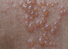
Herpes zoster (HZ, also called shingles) is a painful condition that produces a maculopapular and vesicular rash. Usually, the rash appears along a single dermatome (band) around one side of the body or face. In most cases, pain, tingling, burning, or itching occurs a few days before the rash. Next, blisters form, scabbing over in 7 to 10 days. In…
read more

Frequently asked questions about support surfaces

The National Pressure Ulcer Advisory Panel (NPUAP) describes support surfaces as “specialized devices for pressure redistribution designed for management of tissue loads, microclimate, and/or other therapeutic functions.” These devices include specialized mattresses, mattress overlays, chair cushions, and pads used on transport stretchers, operating room (OR) tables, examination or procedure tables, and gurneys. Some support surfaces are part of an integrated…

Herpes zoster: Understanding the disease, its treatment, and prevention

Herpes zoster: Understanding the disease, its treatment, and prevention

Herpes zoster (HZ, also called shingles) is a painful condition that produces a maculopapular and vesicular rash. Usually, the rash appears along a single dermatome (band) around one side of the body or face. In most cases, pain, tingling, burning, or itching occurs a few days before the rash. Next, blisters form, scabbing over in 7 to 10 days. In…

Knowing when to ask for help

As a wound care expert, you’re probably consulted for every eruption, scrape, and opening in a patient’s skin. Occasionally during a patient assessment, you may scratch your head and ask yourself, “What is this? I’ve never seen anything like it.” Most wound care experts want to help heal everyone, and most of us love a challenge. But when should we…

Managing chronic venous leg ulcers — what’s the latest evidence?

Managing chronic venous leg ulcers — what’s the latest evidence?

Chronic venous leg ulcers (CVLUs) affect nearly 2.2 million Americans annually, including an estimated 3.6% of people over the age of 65. Given that CVLU risk increases with age, the global incidence is predicted to escalate dramatically because of the growing population of older adults. Annual CVLU treatment-related costs to the U.S. healthcare system alone are upwards of $3.5 billion,…

Ostomy documentation tips

General characteristics Document if the diversion is an intestinal or urinary ostomy, whether it’s temporary or permanent, and the location— abdominal quadrant, skin fold, umbilicus. (See Descriptor reference.)

Skin substitutes: Understanding product differences

Skin substitutes (also called tissuebased products and dermal replacements) are a boon to chronic wound management when traditional therapies have failed. When selecting skin substitutes for their formularies, wound care professionals have many product options—and many decisions to make. Repair of skin defects has been a pressing concern for centuries. As early as the 15th century BC, Egyptian physicians chronicled…

2016 Journal: November – December Vol. 5 No. 6

Click here to access the digital edition

Read More

Lymphedema and lipedema: What every wound care clinician should know

Imagine you have a health condition that affects your life every day. Then imagine being told nothing can be done about it; you’ll just have to live with it. Or worse yet, your physician tells you the problem is “you’re just fat.”

Many people with lymphedema or lipedema have no idea their condition has a name or that many other people suffer from the same thing. Although lymphedema and lipedema can’t be cured, proper management and resources can help patients cope. This article improves your grasp of these conditions, describes how to recognize and manage them, and explains how to support your patients.

To understand lymphedema and lipedema, first you need to understand how the lymphatic system functions. It makes lymph, then moves it from tissues to the bloodstream. It also plays a major role in the immune system, aiding immune defense. In addition, it helps maintain normal fluid balance by promoting fluid movement from the interstitial tissues back to the venous circulation. (See Lymphatic system: Four major functions.)

If the lymphatic system is impaired from a primary (hereditary or congenital) condition or a secondary problem, lymphedema can result. In this chronic, potentially progressive, and incurable condition, protein-rich fluid accumulates in the interstitial tissues.

Lymphedema basics

Lymphedema occurs in four stages.

Stage 0. During this stage (also called the subclinical or latency stage), transport capacity of the lymphatic system decreases but remains sufficient to manage normal lymphatic loads. Signs and symptomsaren’t evident and can be measured only by sensitive instruments, such as bioimpedance spectroscopy and optoelectronic volumetry. Without such instruments to quantify volume changes, diagnosis may rest on subjective complaints.

In this stage, limited functional reserve of the lymphatic system leads to a fragile balance between subnormal transport capacity and lymphatic loads. Added stress on the lymphatic system (as from extended heat or cold exposure, injury, or infection) may cause progression to stage 1.

Providing appropriate patient information and education, especially after surgery, can dramatically reduce the risk that lymphedema will progress to a more serious stage.

Stage 1. Considered the spontaneously reversible stage, stage 1 is marked by softtissue pliability without fibrotic changes. Pitting can be induced easily. In early stage 1, limb swelling may recede over – night. With proper management, the patient can expect the extremity to decrease to a normal size compared to that of the uninvolved limb. Otherwise, lymphedema is likely to progress to stage 2.

Stage 1 lymphedema may be hard to distinguish from edemas from other causes. Clinicians must rely on the patient history and monitor for swelling resolution with conventional management, such as compression and elevation, or note if swelling persists despite these standard interventions.

Stage 2. Sometimes called the spontaneously irreversible stage, stage 2 is identified mainly from tissue proliferation and subsequent fibrosis (called lymphostatic fibrosis). The fluid component can be removed spontaneously, but removal of the increased tissue proliferation (initially irreversible) takes more time. Tissue proliferation stems from long-standing accumulation of protein-rich fluid; over time, the tissue hardens and pitting is hard to induce. In many cases, swelling volume increases, exacerbating the already compromised local immune defense.

Consequently, infections (particularly cellulitis) are common; these, in turn, increase the volume of the affected area. Proper treatment can reduce volume.

With proper care (complete decongestive therapy [CDT]), lymphedema can stabilize during stage 2. But patients with chronic or recurrent infections are likely to progress to stage 3.

Stage 3. Also called lymphostatic elephantiasis, this stage is marked by further fluid volume increases and progression of tissue changes. Lymphostatic fibrosis becomes firmer and other skin alterations may occur, including papillomas, cysts, fistulas, hyperkeratosis, fungal infections, and ulcers. Pitting may be present. Natural skinfolds deepen (especially those of the dorsum of the wrist or ankle) and, in many cases, cellulitis recurs.

If lymphedema management starts during this stage, reduction can still occur. Even in extreme cases, with proper care and patient adherence to treatment, lymphostatic elephantiasis can be reduced so the leg is a normal or near-normal size.

Assessment and diagnosis

A thorough physical examination is the gold standard for diagnosing lymphedema. A complete patient history, body-systems review, inspection, and palpation can help determine if edema is lymphedema.

Clinically, the only test with proven reliability and validity in diagnosing lymphedema is the Stemmer sign. Fibrotic changes associated with lymphedema can lead to thickened skin over the proximal phalanges of the toes or fingers. If you can’t tent or pinch the skin on the involved extremity, lymphedema is present (a positive Stemmer sign). However, a negative finding (soft, pliable tissue) doesn’t rule out  lymphedema because the condition may be in an early stage, before tissue proliferation and fibrosis have set in.

Management

Although incurable, lymphedema can be managed successfully through CDT. This approach involves proper identification of lymphedema, manual lymph drainage, skin and nail care, patient education, compression, and exercise.

CDT has two phases:

Phase I, the intensive phase, continues until the extremity has decongested or reached a plateau. The clinician provides treatments and educates the patient about all aspects of CDT to prepare him or her for phase II. Phase I can last several weeks to several months depending on lymphedema severity.

Phase II, the maintenance phase, begins once the extremity has decongested or plateaued. This phase still focuses on CDT, but now the patient, not the clinician, is responsible for all care. The goal is to reduce limb size while enabling the patient to become self-sufficient in managing lymphedema. Although CDT can bring significant improvements in limb size, skin quality, and function, patients must remember that phase II continues lifelong. Be sure to provide education about ongoing self-management strategies.

Lipedema: The disease they call “fat”

Lipedema is a painful disorder of fat deposition. Pathologic deposition of fatty tissue (usually below the waist) leads to progressive leg enlargement. Like lymphedema, lipedema is incurable but manageable. Unless managed properly, lipedema can reduce mobility, interfere with activities of daily living, and lead to secondary lymphedema. (See Lipedema stages.)

Lipedema commonly is misdiagnosed as lymphedema. However, lymphedema involves protein-rich fluid, whereas lip edema is a genetically mediated fat disorder. Because lipedema resists diet and exercise, it can lead to psychosocial complications. Lipedema occurs almost exclusively in women; typically, onset occurs between puberty and age 30. One unpublished epidemiologic study puts lip edema incidence in females at 11%. Some patients have a combination of lipedema and lymphedema. (See Viewing lipolymphedema.)

Assessment and diagnosis

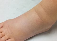
As with lymphedema, lipedema diagnosis rests on clinical presentation. Lipedema characteristics include bilateral and symmetrical involvement, absence of pitting (because lipedema isn’t a fluid disorder), soft and pliable skin, and filling of the retromalleolar sulcus (called the fat pad sign.)

Key signs and symptoms include:

• feeling of heaviness in the legs (aching dysesthesia)

• easy bruising

• sensitivity to touch (called “painful fat syndrome”)

• orthostatic edema

• oatmeal-like changes to skin texture.

Nearly half of lipedema patients are overweight or obese, but many appear of normal weight from the waist up. Essentially, the upper and lower extremities don’t match. The lower extremities typically show fatty deposits extending from the iliac crest to the ankles, sparing the feet. (See Lipedema patterns.)

Management

Lipedema is best  managed through weight control, as additional weight gain through adipose tissue tends to deposit in the legs. For patients with concomitant lymphedema (lipolymphedema), modified CDT helps reduce and manage lymphatic compromise. To address excess fat deposition, newer “wet” liposuction techniques have proven beneficial. These techniques gently detach adipose cells from the tissue, helping to preserve connective tissue and lymphatic vessels.

Know what to look for

In both lymphedema and lipedema, early identification and proper diagnosis are key. (See Differentiating lymphedema and lipedema.) A thorough history and physical exam will likely lead to an accurate diagnosis, if clinicians know what to look for. Proper diagnosis and treatment can prevent expensive and ineffective interventions, which can negatively affect both the patient’s condition and psychological well being.

Heather Hettrick is an associate professor at Nova Southeastern University, Department of Physical Therapy in Fort Lauderdale, Florida.

Selected references

Fat Disorders Research Society. Lipedema description.

Fife CE, Maus EA, Carter MJ. Lipedema: a frequently misdiagnosed and misunderstood fatty deposition syndrome. Adv Skin Wound Care. 2010;23(2):81-92

Herbst KL. Rare adipose disorders (RADS) masquerading as obesity. Acta Pharmacol Sin. 2012;33(2):155-72.

Lipedema Project.

National Lymphedema Network. Position papers.

Schmeller W, Hueppe M, Meier-Vollrath I. Tumescent liposuction in lipoedema yields good long-term results. Br J Dermatol. 2012;166(1):161-8.

Zuther J. A closer look at lipedema and the effects on the lymphatic system. December 13, 2012. lymphedemablog.com/2012/12/13/a-closer-look-at-lipedema-and-the-effects-on-the-lymphatic-system/

Zuther J. Stages of lymphedema. October 3, 2012.

Read More

Assessing footwear in patients with diabetes

Inappropriate footwear is the most common source of trauma in patients with diabetes. Frequent and proper assessment of appropriate footwear is essential for protecting the diabetic foot from ulceration.

Here is a step-by-step process for evaluating footwear. Be sure to evaluate footwear with the patient walking, standing, and sitting. (more…)

Read More

Resource Center

Infographic: An Average Day in the Life of Nursing

Opioid Addiction by the Numbers

Quizzes

Quizzes
– Wound Infections
– Hyperbaric Oxygen Therapy
– Assess Diabetic Foot
– Ram’s Horn

Read More

How to apply silver nitrate

Topical application of silver nitrate is often used in wound care to help remove and debride hypergranulation tissue or calloused rolled edges in wounds or ulcerations. It’s also an effective agent to cauterize bleeding in wounds. Silver nitrate is a highly caustic material, so it must be used with caution to prevent damage to healthy tissues. (more…)

Read More

No more skin tears

Imagine watching your skin tear, bleed, and turn purple. Imagine, too, the pain and disfigurement you’d feel.

What if you had to live through this experience repeatedly? That’s what many elderly people go through, suffering with skin tears through no fault of their own. Some go on to develop complications.

A skin tear is a traumatic wound caused by shear, friction, or blunt-force trauma that results in a partial- or full-thickness injury. Skin tears are painful because the precipitating injury commonly involves the dermis, which is rich with nerve endings. (more…)

Read More
1 2 3 4 5 8