Wounds on the Web: Accessing the best online resources

By Donna Sardina, RN, MHA, WCC, CWCMS, DWC, OMS

Knowledge is exploding online, making it essential that you’re comfortable using the Internet. You can also go online to save time and find a job, among other tasks. (See Online value.)

However, you also need to keep in mind that anyone can put information on the Internet. As the caption of a cartoon by Peter Steiner, published in The New Yorker says, “On the Internet, nobody knows you’re a dog.” (more…)

Read More

Clinician Resources


Here are resources that can help you in your busy clinical practice by giving you information quickly. Don’t miss out!

Free educational and clinical resources on pressure ulcers

Access valuable educational and clinical resources from the National Pressure Ulcer Advisory Panel. Among the free resources are:
• poster of best practices for prevention of medical device–related pressure ulcers (available as a PDF)
• pressure-ulcer prevention points (available as a PDF)
registered nurse competency-based curriculum: pressure ulcer prevention.

Tutorials on diabetes foot care

Take an interactive tutorial on foot care for patients with diabetes. The module was published by the Patient Education Institute. You can also choose to watch in a self-playing mode and download a PDF text summary.

Quality assurance resources for long-term care

The resources in “Quality Assurance Performance Improvement (QAPI) Resources” are targeted toward those working in long-term care. The list includes links such as:
Advancing Excellence in America’s Nursing Homes National Campaign, which has resources and tools for nursing homes to improve in several different areas
Implementing Change in Long-Term Care, a detailed guide on how to engage nursing home staff in the change process
The Long-Term Care Improvement Guide, developed by Planetree in partnership with Picker Institute.

Prescription assistance programs

Do you have patients who need help paying for their prescriptions? “Understanding Prescription Assistance Programs,” from the National Council on Patient Information and Education, explains how these programs work and provides resources.

Read More

From the Editor – Wound care superhero

by Donna Sardina, RN, MHA, WCC, CWCMS, DWC, OMS

What an honor it is to be the wound care “superhero”—the guru, the healer, the go-to person. Unfortunately, this honor may be accompanied by wound care overload—too much to do in too little time.

Once someone is crowned the superhero specialist, others may try to transfer every aspect of wound and skin care to that person—all treatment plans, assessments, documentation, prevention, education, and accountability. Superheroes don’t cry, so they don’t complain about the workload. Yet, the overload must be controlled. (more…)

Read More

Understanding stoma complications

By Rosalyn S. Jordan, RN, BSN, MSc, CWOCN, WCC, OMS; and Judith LaDonna Burns, LPN, WCC, DFC

About 1 million people in the United States have either temporary or permanent stomas. A stoma is created surgically to divert fecal material or urine in patients with GI or urinary tract diseases or disorders.

A stoma has no sensory nerve endings and is insensitive to pain. Yet several complications can affect it, making accurate assessment crucial. These complications may occur during the immediate postoperative period, within 30 days after surgery, or later. Lifelong assessment by a healthcare provider with knowledge of ostomy surgeries and complications is important. (more…)

Read More

Compression therapy for chronic venous insufficiency, lower-leg ulcers, and secondary lymphedema

By Nancy Chatham, RN, MSN, ANP-BC, CCNS, CWOCN, CWS, and Lori Thomas, MS, OTR/L, CLT-LANA

An estimated 7 million people in the United States have venous disease, which can cause leg edema and ulcers. Approximately 2 to 3 million Americans suffer from secondary lymphedema. Marked by abnormal accumulation of protein-rich fluid in the interstitium, secondary lymphedema eventually can cause fibrosis and other tissue and skin changes. (more…)

Read More

What’s causing your patient’s lower-extremity redness?

patient lower extremity redness

By Robyn Bjork, MPT, CWS, WCC, CLT-LANA

The ability to understand or “read” lower-extremity redness in your patient is essential to determining its cause and providing effective treatment. Redness can occur in multiple conditions—hemosiderin staining, lipodermatosclerosis, venous dermatitis, chronic inflammation, cellulitis, and dependent rubor. This article provides clues to help you differentiate these conditions and identify the specific cause of your patient’s lower-extremity redness. (more…)

Read More

Apply QAPI to reduce pressure ulcer rates

By Jeri Lundgren, BSN, RN, PHN, CWS, CWCN

The Affordable Care Act of 2010 requires nursing homes to have an acceptable Quality Assurance and Performance Improvement (QAPI) plan within a year after the start of the QAPI regulation. While the implementation of this regulation may be a year out, now is the time to start applying its principles. Reducing pressure ulcer rates is a great program to target for a QAPI plan.

A team approach

If you decide to use pressure ulcers as your QAPI project, don’t take on your entire program at once. Break the program down into system subsets (for example, admission process, prevention program, and weekly rounds). Determining the status of your program in each subset—completed, needs improvement, or not completed—can help you prioritize which areas to target. It’s important you have support from leadership for your efforts.

I’ll use the example of the admission phase (ensuring that within the first 24 hours, skin and risk concerns are identified and a temporary plan of care is implemented) to illustrate a QAPI project. To address this area, a team was created, including representation from staff members involved with the admission process. The team then used the problem-solving model Plan-Do-Study-Act (PDSA) to examine the process.

The first step in the PDSA cycle is to Plan. During this step, you:

• evaluate and analyze the current process to determine baseline data, which are used to measure progress
• identify system performance gaps
• determine the root cause of the performance gaps
• develop an action plan that identifies the goals, steps, responsible staff, and target dates.

In our example, the team determined that within the first 24 hours, skin inspections were being completed only 10% of the time. The root-cause analysis revealed that the admission nurses didn’t feel competent to document identified pressure ulcers or skin concerns, so they deferred it until the wound nurse was available. The team’s action plan included the following:

• Develop and educate all the facility nurses on how to complete and accurately document a skin inspection.
• Develop and implement a competency evaluation to assess the nurse’s ability to apply the knowledge at the bedside.
• Develop an ongoing plan to ensure all nurses receive this education during orientation and yearly thereafter.

The team also set the following goal:
By the end of the next quarter, 100% of admitted patients will have an accurate skin inspection completed within 24 hours of admission.

The second step of the PDSA cycle is Do. During this step, you implement and execute the plan, while documenting your observations and recording data.

In our example, the “Do” was to:
• develop and provide the skin inspection education and bedside competency evaluations
• develop an evaluation and tracking
system
• add the education to the orientation program
• add the education to the staff development calendar to be offered yearly.

The third step of the PDSA cycle is to Study: In this phase, you:
• reevaluate and analyze the system
• compare the results with the baseline data and predictions
• summarize what was learned and accomplished and what needs to be improved
• determine if another PDSA cycle is
necessary to continue to improve the system.

Once all staff had been properly educated and competency testing completed, an analysis of the rate and accuracy of the admission skin inspections done within 24 hours of admission was completed. It was found that 100% of the patients admitted had a complete skin inspection done within 24 hours. However, not all the nurses could accurately stage pressure ulcers, so it was determined that the system needed improvement to ensure accurate assessments.

The last step of the PDSA cycle is to Act. In this step, you:
• determine what changes need to be made
• modify the plan to continue to improve the system
• repeat the PDSA cycle as necessary.

In our example, the team determined the nurses needed more guidance and education on staging of pressure ulcers. Therefore, a new PDSA cycle was set to ensure the nurses are competent in this area.

Benefits for staff and patients

It may be difficult to start the QAPI project and at times the process may be stressful, but keep in mind that a successful pressure ulcer QAPI project can improve not only the quality of life and care of your patients but also morale and team building for your staff. n

Jeri Lundgren is director of clinical services at Pathway Health in Minnesota. She has beenspecializing in wound prevention and management since 1990.


Read More

Clinical Notes

Study finds less-invasive method for identifying osteomyelitis is effective

Researchers have found that using hybrid 67Ga single-photon emission computed tomography and X-ray computed tomography (SPECT/CT) imaging combined with a bedside percutaneous bone puncture in patients with a positive scan is “accurate and safe” for diagnosing osteomyelitis in patients with diabetes who have a foot ulcer without signs of soft-tissue infection.

The new method, which avoids an invasive bone biopsy, has a sensitivity of 88% and a specificity of 93.6%. In the study of 55 patients, antibiotic treatment was avoided in 55% of suspected cases.

Diagnosing diabetic foot osteomyelitis in patients without signs of soft tissue infection by coupling hybrid 67Ga SPECT/CT with bedside percutaneous bone puncture,” published by Diabetes Care, followed patients for at least a year.

MRSA strains will likely continue to coexist in hospitals and communities

The strains of methicillin-resistant Staphylococcus aureus (MRSA) differ in the hospital and community settings, and both are likely to coexist in the future, according to a study in PLOS Pathogens.

Hospital-community interactions foster coexistence between methicillin-resistant strains of Staphylococcus aureus” notes that previously it was thought that the more invasive community strains would become more prevalent (and even eliminate) hospital strains. This new information could have significant consequences for public health because of the differences in the resistance of the two strains.

C. difficile prevention actions fail to stop spread

Despite increasing activities to prevent the spread of Clostridium difficile, infection from C. difficile remains a problem in healthcare facilities, according to a survey of infection preventionists by the Association for Professionals in Infection Control and Epidemiology (APIC).

The survey found that 70% of preventionists have adopted additional interventions in their healthcare facilities since March 2010, but only 42% have seen a decline in C. difficile infection rates; 43% saw no decline.

A total of 1,087 APIC members completed the survey in January 2013. The survey also found that more than 92% of respondents have increased emphasis on environmental cleaning and equipment decontamination practices, but 64% said they rely on observation, rather than more accurate and reliable monitoring technologies, to assess cleaning effectiveness.

In addition, 60% of respondents have antimicrobial stewardship programs at their facilities, compared with 52% in 2010. Such programs promote the appropriate use of antibiotics, which can help reduce the risk of C. difficile infection.

According to the Centers for Disease Control and Prevention, diarrhea caused by C. difficile is linked to 14,000 American deaths each year.

Mast cells may not play significant role in wound healing

Evidence that mast cells are not required for healing of splinted cutaneous excisional wounds in mice,” published in PLOS One, analyzed wound healing in three types of genetically mast-deficient mice and found they reepithelialized their wounds at rates similar to control mice. At the time of closure, the researchers found that scars in all the mice groups were similar in both “quality of collagen deposition and maturity of collagen fibers.” The findings fail to support the previously held belief that mast cells are important in wound healing.

Study identifies effective casting for diabetes-related plantar foot ulcers

Nonremovable casts that relieve pressure are more effective than removable casts or dressings alone for the treatment of plantar foot ulcers caused by diabetes, according to an analysis of clinical trials.

The authors of “Pressure-relieving interventions for treating diabetic foot ulcers,” published by The Cochrane Library, reviewed 14 randomized clinical trials that included 709 participants. Nonremovable pressure-relieving casts were compared to dressings alone, temporary therapeutic shoes, removable pressure-relieving devices, and surgical lengthening of the Achilles tendon.

The study also notes that when combined with Achilles tendon lengthening, nonremovable devices were more successful in one forefoot ulcer study than the use of a nonremovable cast alone.

Most studies were from the United States (five) and Italy (five), with Germany, the Netherlands, Australia, and India each contributing one study.

Prescriber preference drives use of antibiotics in long-term care

Prolonged antibiotic treatment in long-term care: Role of the prescriber,” published by JAMA Internal Medicine, found that the preferences of prescribers, rather than patient characteristics, drive antibiotic treatment.

The study of 66,901 patients from 630 long-term care facilities found that 77.8%
received a course of antibiotics. The most common length (41%) was 7 days, but the length exceeded 7 days in 44.9% of patients. Patient characteristics were similar among short-, average-, and long-duration prescribers.

The study authors conclude: “Future trials should evaluate antibiotic stewardship interventions targeting prescriber preferences to systematically shorten average treatment durations to reduce the complications, costs, and resistance associated with antibiotic overuse.”

Electrophysical therapy may be helpful for diabetic foot ulcers

Electrophysical therapy for managing diabetic foot ulcers: A systematic review” concludes that electrophysical therapy is potentially beneficial because in each randomized clinical trial it outperformed the control or sham electrical stimulation.

The authors of the study in International Wound Journal reviewed eight trials with a combined total of 325 patients. Five studies were on electrical stimulation, two on phototherapy, and one on ultrasound. Because of the small number of trials, the possibility of harmful effects can’t be ruled out, and the authors recommend “high-quality trials with larger sample sizes.”

Significant geographic variations in spending, mortality exist for diabetic patients with foot ulcers and amputations

Geographic variation in Medicare spending and mortality for diabetic patients with foot ulcers and amputations” reports healthcare spending and mortality rates vary “considerably” across the United States.

The study in Journal of Diabetes and Its Complications found that higher spending wasn’t associated with a significant reduction in 1-year patient mortality. In addition, rates of hospital admission were associated with higher per capita spending and higher mortality rates for patients.

Home-based exercise program improves life for lymphedema patients

An individualized, home-based progressive resistance exercise program improves upper-limb volume and circumference and quality of life in postmastectomy patients with lymphedema, according to a study published in the Journal of Rehabilitation Research and Development.

Effect of home-based exercise program on lymphedema and quality of life in female postmastectomy patients: Pre-post intervention study” included 32 women who participated in an 8-week program. The women received education about the program and an initial training session from a physiotherapist. They practiced the exercise sequence and received a program and logbook once their performance was satisfactory. Patients were told to increase weight only when two sets of 15 repetitions became easy to perform.

Read More

Is your wound-cleansing practice up to date?

wound cleansing practice

By Donna Sardina, RN, MHA, WCC, CWCMS, DWC, OMS

With so much focus on dressing choices, it’s easy to forget the importance of wound cleansing. Cleaning a wound removes loose debris and planktonic (free-floating) bacteria, provides protection to promote an optimal environment for healing, and facilitates wound assessment by optimizing visualization of the wound. You should clean a wound every time you change a dressing, unless it’s contraindicated.

Here’s a review of how to choose and use a wound cleanser so you can see if your practice is up to date. (more…)

Read More

What you need to know about hydrocolloid dressings

hydrocolloid dressing example1

By Nancy Morgan, RN, BSN, MBA, WOC, WCC, DWC, OMS

Each month, Apple Bites brings you a tool you can apply in your daily practice.

Description

A hydrocolloid dressing is a wafer type of dressing that contains gel-forming agents in an adhesive compound laminated onto a flexible, water-resistant outer layer. Some formulations contain an alginate to increase absorption capabilities. The wafers are self-adhering and available with or without an adhesive border and in various thicknesses and precut shapes for such body areas as the sacrum, elbows, and heels. Click here to see examples of
hydrocolloid dressings.

(more…)

Read More

Stand up to bullies

By: Donna Sardina, RN, MHA, WCC, CWCMS, DWC, OMS

At some point, most of us have encountered a bully—most commonly when we were kids. You might think that as we get older, bullying wouldn’t be a problem we have to deal with. Unfortunately, that’s not the case. In the healthcare field, bullying can be even worse than it was when we were children.

Bullying in health care takes many different forms, including fighting among different types of clinicians, managers bullying subordinates, peer-to-peer bullying and, most commonly, specialists bullying other specialists. Years ago when I realized my dream of becoming a wound care specialist, I thought other specialists would be relieved I was on board to help with the overwhelming task of spreading wound care knowledge and healing wounds. But I found out quickly that I was pretty much alone with those thoughts, and my first encounter with wound care bullies occurred.

I began to ask myself: What did I do wrong? Why are they slamming me? What did I do to them? They don’t even know me; they’ve never even talked to me. This may sound familiar to many of you, whether you’re a wound care specialist, an ostomy specialist, or a diabetes or lymphedema specialist.

Workplace bullying is defined as repeated, unreasonable actions by individuals (or a group) directed toward an employee (or group of employees) that are intended to intimidate, degrade, humiliate, or undermine. Bullying occurs for many reasons; these reasons almost always include insecurity, competition, and the desire to feel more powerful and be in control.

So how do we deal with the bullies?
• Follow the Golden Rule: Treat others as you’d like others to treat you. Don’t stoop to the bully’s level.
• Stay calm and rational. Don’t get emotional. Bullies take pleasure in manipulating people emotionally.
• Don’t lose your confidence or blame yourself. Recognize that this isn’t about you; it’s about the bully. Be proud and confident in your certification credential.
• Focus on your purpose—to provide safe, competent, high-quality care to every patient.
• Document the bullying incident. Start a diary detailing the nature of the bullying, including dates, times, places, what was said or done, and who was present. Start a file with copies of anything in print that shows harassment and bullying; hold onto copies of documents that contradict the bully’s accusations against you.
• If the bullying behavior compromises patient safety and care, report the bully.

Stopping all bullying in health care may seem like an insurmountable goal, but I believe that together we can try to stop the bullying cycle in our specialty. By setting the example and supporting each other, we can turn the focus back to healing and caring for our patients as a team, not as one practitioner against the world.

Actions speak louder than words. As Ralph Waldo Emerson said, “What you do speaks so loudly that I cannot hear what you say.”

Donna Sardina, RN, MHA, WCC, CWCMS, DWC, OMS
Editor-in-Chief
Wound Care Advisor
Cofounder, Wound Care Education Institute
Plainfield, Illinois

Read More

Understanding peristomal skin complications

By Rosalyn Jordan, RN, BSN, MSc, CWOCN, WCC, and Marci Christian, BBE

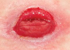
Any patient with a fecal or urinary ostomy may experience complications on the skin surface around the stoma. These complications may occur lifelong, although they’re more common during the first 5 years after the initial ostomy surgery. Causative factors include infection, trauma, certain diseases, and chemical irritation; most of these problems stem from the pouching system or pouch leakage.

Peristomal skin complications can cause a wide range of signs and symptoms, from skin discoloration to polyp-like growths, from erythema to full-thickness wounds. They can lead to discomfort, pain, poor self-image, social isolation, and impaired quality of life, not to mention additional care costs.

Incidence and types of these complications are hard to compare or contrast across multiple patients. Until recently, no standardized assessment or documentation tools were available to characterize or define complications. For this reason, reported rates ranged widely, from 10% to 70%. And because no designated common language or categories related to peristomal skin complications existed, documentation was inconsistent.

Download “How to Use” education program for the Ostomy Skin Tool

Ostomy Skin Tool

In the late 2000s, a group of nurses experienced in caring for ostomy patients worked with the World Council of Enterostomal Therapists to develop a resource called the Ostomy Skin Tool, which clinicians can use to categorize and describe peristomal skin complications in a consistent, objective manner. The tool also provides a common language for documentation.

The Ostomy Skin Tool has three major assessment domains—discoloration (D), erosion/ulceration (E), and tissue overgrowth (T), known collectively as DET. The DET combined rating ranges from normal, rated 0, to the worst condition possible, rated 15. Mild DET complications are documented as less than 4, moderate as less than 7, and severe as 8 or higher. (See Using the Ostomy Skin Tool by clicking the PDF icon above.)

The tool describes four categories of peristomal complications:
• chemical irritation
• mechanical trauma
• disease-related complications
• infection-related complications.

Chemical irritation

Chemical irritation can stem from irritants (as in contact dermatitis) or allergic reactions (allergic dermatitis). The most likely cause of chemical dermatitis is effluent leakage (feces or urine) from the colostomy, ileostomy, or urostomy, in which effluent comes in contact with peristomal skin. Other potential causes include contact with soap, certain adhesives, and adhesive removers.

The major treatment of chemical irritation is identification and removal of the offending agent, followed by patient and caregiver education on the new pouching procedure the patient must use. Follow-up assessment also is recommended. In a 2010 study that followed 89 patients for 1 year after ostomy surgery, about 50% of subjects experienced peristomal skin complications, most of them from pouch leakage. Another investigator estimated that 85% of ostomy patients experience pouch leakage at some time during their lives. Pouch leakage usually occurs when stool is extremely liquid (for instance, ileostomy effluent). Other causes of pouch leakage include wearing a pouch more than half full of effluent and abdominal contours that aren’t level. Besides changes in the pouching system, treatment may entail adding products to the pouching system or removing certain agents.

Some patients experience allergic dermatitis in reaction to products used in the pouching system (such as skin barriers, belts, pouch closures, or adhesives). However, allergic dermatitis is rare. One 2010 study suggested allergic reactions to these products occur in only about 0.6% of patients with peristomal skin irritation. Most major ostomy product manufacturers provide a patch test on request to help identify allergic conditions. Once the offending product is discontinued, allergic dermatitis should resolve rapidly.

Mechanical trauma

Mechanical trauma usually results from either the pouching system itself or its removal. It also may result from harsh or multiple skin-barrier removals, pressure from convex rings or pouches, and abrasive cleansing techniques. Some researchers believe the stronger the adhesive barrier and the more often a pouch is changed, the greater the risk of epidermal damage.

Mechanical trauma may present as a partial-thickness ulcer caused by pressure, shear, friction, tearing, or skin stripping. Patients with fragile skin are susceptible to mechanical trauma, so less aggressive pouching systems may be preferred for them. Of course, if the pouching system is changed, the patient or caregiver needs to learn about the new system.

Disease-related complications

Disease-related peristomal complications may be linked to preexisting skin conditions, such as psoriasis, eczema (atopic dermatitis), or seborrheic dermatitis. Hyperplasia also may occur. This overgrowth of cells, which may appear as gray or reddish brown pseudoverrucous lesions, usually is linked to urinary ostomies, although it can occur with fecal ostomies as well. Vinegar soaks are the recommended treatment, in addition to a change in the pouching system and corresponding patient education.

Occasionally, other disease-related complications occur, including primary adenocarcinoma of the peristomal skin and peristomal pyoderma gangrenosum, a painful and problematic condition that presents as peristomal ulcers. Ulcer borders are well-defined with a bluish purple coloration at the edges. Infection must be ruled out, as this condition usually is linked to an autoimmune condition. Treatment includes pain management and, in most cases, a topical corticosteroid. Crohn’s disease also may manifest as a peristomal skin ulcer.

Infection-related complications

Infection-related complications may be bacterial or fungal. Two common peristomal skin infections are folliculitis and Candida fungal infections. An infection of the hair follicle that causes pustules, folliculitis usually stems from traumatic hair pulling in the peristomal area during pouch removal. It may warrant a prescribed antibiotic, along with patient teaching regarding proper hair removal using an electric razor.

Candida infections may arise because peristomal skin provides a warm, dark, moist environment that promotes fungal growth. These infections appear as erythema with pustules or papules and satellite lesions. Treatment usually involves antifungal powder and use of the crusting technique to secure the pouching system. (See Using the crusting technique by clicking the PDF icon above.)

Management

Many complications are well advanced by the time patients seek assistance, perhaps because they don’t understand the significance of their symptoms and think they can manage the problem themselves. In some cases, they don’t know where to turn for assistance. Commonly, the complication progresses to the point where the patient goes to the emergency department or (particularly during the immediate postoperative period) needs to be readmitted for treatment. The best way to manage peristomal skin complications is to prevent them in the first place. (See Preventing peristomal skin complications by clicking the PDF icon above.)

Patient education

Over the past 20 years, hospital stays for ostomy surgery patients have decreased from about 2 weeks to less than 5 days. Reduced stays decrease the time available for caregivers to teach patients and family members how to empty and change the pouch. They need alternative education covering (among other topics) how to recognize peristomal skin complications and when to seek help. Not only do these complications require vigilant self-observation, but many patients don’t understand their implications or how rapidly they can worsen. In some cases, the first symptoms are itching and redness under the skin barrier. Fortunately, some patients may know or remember that itching, burning, stinging, reddened, or weeping peristomal skin requires professional attention. They can avoid serious complications by seeking assistance early, such as right after noticing pouch leakage.

Early treatment can reduce the cost of treatment. In a 2012 study, researchers estimated care costs related to peristomal skin complications for a 7-week treatment period, using the Ostomy Skin Tool as a reference. Severe complications (those with a DET score above 8) cost six times more to treat than mild cases (those with a DET score below 4) and 4.5 times more than moderate cases.

Along with early intervention by a trained ostomy care specialist, self-assessment by ostomy patients promotes a better quality of life, reduces pain, and may decrease care costs. Clinicians’ use of the Ostomy Skin Tool to assess and document peristomal skin complications promotes more reliable, objective, comparable assessment data for reporting.

Selected references
Al-Niaimi F, Lyon CC. Primary adenocarcinoma in peristomal skin: a case study. Ostomy Wound Manage. 2010;56(1):45-7.

Burch J. Management of stoma complications. Nurs Times. 2011;107(45):17-8, 20.

Jemec GB, Martins L, Claessens I, et al. Assessing peristomal skin changes in ostomy patients: validation of the Ostomy Skin Tool. Br J Dermatol. 2011; 164;330-5.

Jones T, Springfield T, Brudwick M, Ladd A. Fecal ostomies: practical management for the home health clinician. Home Healthc Nurse. 2011;29(5):306-17.

Martins L, Samai O, Fernandez A, et al. Maintaining healthy skin around an ostomy: peristomal skin disorders and self-assessment. Gastrointest Nurs. 2011;
9(2):9-13.

Martins L, Tavernelli K, Serrano JLC. Introducing a peristomal skin assessment tool: The Ostomy Skin Tool. World Council Enterostomal Therapists J. 2008;28(2):3-13.

Meisner S, Lehur P, Moran B, et al. Peristomal skin complications are common, expensive, and difficult to manage: a population based cost modeling study. PLoS One. 2012;7(5):e37813.

Nybaek H, Jemec GB. Skin problems in stoma patients. J Eur Acad Dermatol Venereol. 2010;24(3):249-57.

Omura Y, Yamabe M, Anazawa S. Peristomal skin disorders in patients with intestinal and urinary ostomies: influence of adhesive forces of various hydrocolloid wafer skin barriers. J Wound Ostomy Continence Nurs. 2010;37(3):289-98.

Ratliff CR. Early peristomal skin complications reported by WOC nurses. J Wound Ostomy Continence Nurs. 2010;37(5):505-10.

Shabbir J, Britton DC. Stomal complications: a literature overview. Colorectal Dis. 2010;12(10):958- 64.

Wound, Ostomy, Continence Clinical Practice Ostomy Subcommittee. Peristomal skin complications: Best practice for clinicians. Mt. Laurel, NJ; 2007.

The authors work for RecoverCare, LLC, in Louisville, Kentucky. Rosalyn Jordan is director of clinical education and Marci Christian is a clinical associate product specialist.

Read More
1 27 28 29 30 31 37