By Robyn Bjork, MPT, CWS, WCC, CLT-LANA
The ability to understand or “read” lower-extremity redness in your patient is essential to determining its cause and providing effective treatment. Redness can occur in multiple conditions—hemosiderin staining, lipodermatosclerosis, venous dermatitis, chronic inflammation, cellulitis, and dependent rubor. This article provides clues to help you differentiate these conditions and identify the specific cause of your patient’s lower-extremity redness.
Hemosiderin staining and lipodermatosclerosis
Hemosiderin staining is dark purple or rusty discoloration of the lower legs caused by chronic venous disease. A 2010 study found hemosiderin staining in all subjects with lipodermatosclerosis and venous ulcers. When vein valves fail, regurgitated blood forces red blood cells (RBCs) out of capillaries. Dead RBCs release iron, which is stored in tissues as hemosiderin, staining the skin.
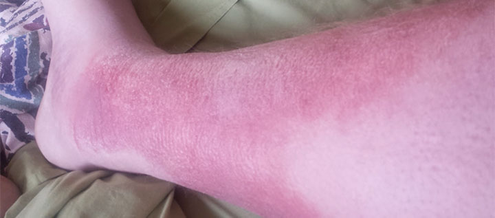
Hemosiderin staining and active lipodermatosclerosis may be misdiagnosed as cellulitis. Active lipodermatosclerosis causes painful, sharply demarcated red patches on medial aspects of the lower leg. Unlike in cellulitis, redness in lipodermatosclerosis is localized to areas of hemosiderin staining and induration. Also, the skin isn’t hot and the patient is afebrile and unresponsive to antibiotics. Lipodermatosclerosis progresses to fibrosis and constriction, causing an inverted champagne-bottle appearance of the legs.
Treat active lipodermatosclerosis with compression therapy and topical corticosteroids, if needed. Control chronic venous hypertension with compression, and hemosiderin staining will fade. Refer the patient for potential corrective venous surgical procedures.
Venous dermatitis
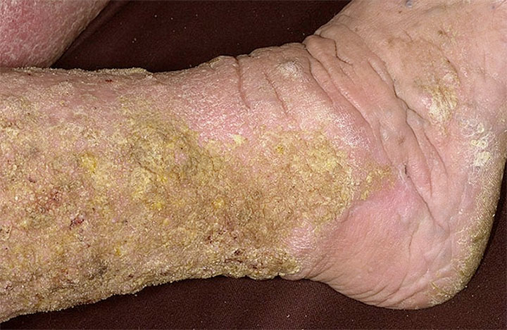
Defined as inflammation of the epidermis and dermis, venous (stasis) dermatitis is common in patients with lower-extremity venous disease. Signs and symptoms include scaling, crusting, weeping, erythema, erosions, and intense itching. This disorder increases the risk of contact sensitivity. Advise the patient to avoid such products as lanolin, balsam of Peru, rubber, adhesives, fragrances, dyes, preservatives, skin sealants, silver sulfadiazine, neomycin, and bacitracin—all known to exacerbate venous dermatitis.
Venous dermatitis commonly is confused with cellulitis. A 2011 study found that 28% of 145 patients hospitalized for cellulitis had been misdiagnosed. The most common mistaken diagnosis was venous dermatitis. Unlike cellulitis, venous dermatitis causes itching and crusting; also, the skin isn’t acutely painful or hot and the patient is afebrile.
Treat acute venous dermatitis with compression therapy and mild-potency topical corticosteroids. Apply corticosteroids sparingly to affected areas once or twice daily for 2 weeks; be aware that premature discontinuation can lead to recurrence, while prolonged use can cause skin thinning and reduced efficacy. Domeboro soaks also decrease weeping, irritation, and itching. Paste bandages impregnated with calamine or zinc oxide are soothing and drying. However, some patients may react to the preservatives in paste bandages, so a patch test is prudent.
Chronic inflammation
Lymphedema causes chronic inflammation. About 50% of plasma proteins leak into the interstitial space daily and are recycled through the lymphatics. Lymphatic failure traps proteins in the tissues; the proteins act like sponges, attracting and binding fluid. The proteins then denature, triggering a chronic inflammatory response. This response sometimes is misdiagnosed and treated as chronic cellulitis.
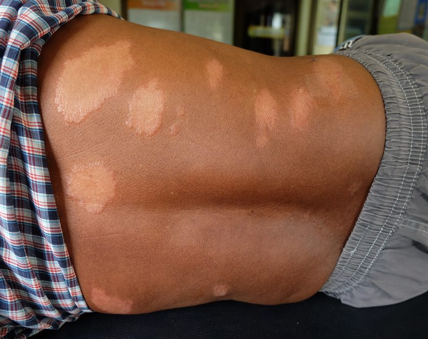
Compared to cellulitis, high-protein chronic inflammation is diffuse and nontender, with light redness and mildly increased warmth. Local skin changes may include thickening or papillomatosis (a lumpy, bumpy appearance). A positive Stemmer’s sign confirms lymphedema. Complete decongestive physiotherapy promotes protein reabsorption and resolves chronic inflammation.
Cellulitis
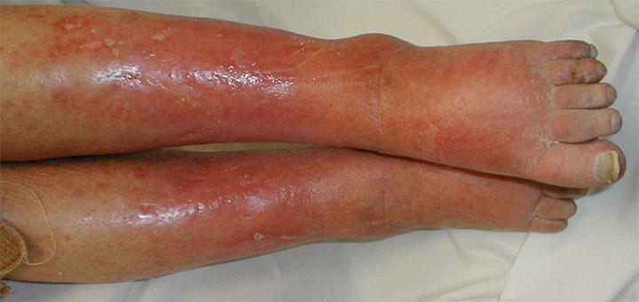
Cellulitis is a rapidly spreading infection of the dermis and subcutaneous tissue. In adults, it most commonly stems from Staphylococcus aureus infection of the legs. Erysipelas, a superficial form of cellulitis, involves the lymphatic system and is differentiated by “streaking” toward a regional lymph node.
Cellulitic skin is hot, acutely painful, edematous, and indurated. Redness spreads and the borders usually are irregular, sharply defined, and slightly elevated. Blisters, hemorrhagic bullae, abscesses, erosions, and necrosis may develop. About 30% to 80% of patients with lower limb cellulitis are afebrile. The white blood cell count, erythrocyte sedimentation rate, and C-reactive protein levels commonly are elevated, but normal values don’t rule out cellulitis.
Treat cellulitis with oral antibiotics effective against staphylococcus and streptococcus. Adding a brief course of oral corticosteroids significantly shortens cellulitis duration. Severe cases may necessitate hospitalization and I.V. antibiotics, plus abscess incision and drainage. Control edema with bed rest and leg elevation.
Recurrent cellulitis is common in patients with lymphedema. With compromised skin immunity, bacteria invade and spread with little resistance. If lymphedema is present, refer the patient for treatment after acute cellulitis resolves. If the patient already is being treated for lymphedema, suspend manual lymphatic drainage and compression until acute cellulitis resolves.
The most common disorders mistaken for lower-limb cellulitis are venous dermatitis, lipodermatosclerosis, irritant dermatitis, and lymphedema. It also may be mistaken for deep vein thrombosis (DVT) or dependent rubor. Rule out DVT using venous duplex ultrasound. Dependent rubor disappears with leg elevation, whereas cellulitic redness doesn’t.
Dependent rubor
Dependent rubor is a fiery to dusky-red coloration visible when the leg is in a dependent position but not when it’s elevated above the heart. The underlying cause is peripheral arterial disease (PAD), so the extremity is cool to the touch. To test for dependent rubor, position the patient supine and elevate the legs 60 degrees for 1 minute; then examine sole color. PAD causes the soles to change from pink to pale in fair-skinned people and to gray or ashen in dark-skinned people. The faster the pallor appears, the worse the PAD. Pallor within 25 seconds of leg elevation indicates severe occlusive disease, which warrants further evaluation for potential revascularization.
Next, observe skin color changes with the patient in a sitting position. Normally, the foot and leg should remain pink with elevation and dependency. In PAD, the color changes from pale to pink and then progresses to purple-red or bright red. The longer dependent rubor takes to reappear, the worse the PAD. Rubor that appears in 25 to 40 seconds indicates severe ischemia. If rubor disappears quickly with elevation and returns in less than 25 seconds, consider the possibility that the patient has venous reflux, not PAD. In this case, pooled blood causing the rubor drains rapidly from the veins when the leg is elevated and regurgitates back into the tissues when the leg is dependent.
If you detect dependent rubor, obtain the ankle-brachial index (ABI) to confirm PAD. For moderate PAD (ABI of 0.5 to 0.79), refer the patient for a routine vascular specialist consultation. For severe PAD (ABI below 0.5), maintain dry, stable wound eschar and urgently refer the patient to a vascular specialist for potential revascularization.
Click here to view images and read a case study on dependent rubor.
Knowledge summary
“Reading” the common causes of leg redness helps you determine what’s causing your patient’s redness so you can provide effective treatment. Remember—chronic venous disease causes hemosiderin staining, lipodermatosclerosis, and venous dermatitis. Dermatitis is itchy and crusty; lipodermatosclerosis causes sclerosis and an inverted champagne-bottle appearance of the legs. Relieve inflammation and itching with topical corticosteroids and treat venous disease with compression and corrective surgery. Lymphedema causes chronic inflammation; treat with complete decongestive physiotherapy. Cellulitis is a spreading skin infection that’s acutely painful and hot; treat with antibiotics. PAD causes dependent rubor, which disappears with leg elevation.
Selected references
Abbade LP, Lastória S, Rollo Hde A. Venous ulcer: clinical characteristics and risk factors. Int J Dermatol. 2011;50(4):405-11.
Bailey E, Kroshinsky D. Cellulitis: diagnosis and management. Dermatol Ther. 2011;24(2):229-39.
Beasley A. Management of patients with cellulitis of the lower limb. Nurs Stand. 2011;26(11):50-5.
Bryant R, Nix D. Acute and Chronic Wounds:
Current Management Concepts. 4th ed. St. Louis, MO: Mosby; 2011.
Buttaro TM, Trybulski J, Polgar P, Sandberg-Cook, J. Primary Care: A Collaborative Practice. 4th ed. St. Louis, MO: Mosby; 2012.
Caggiati A, Rosi C, Casini A, et al. Skin iron deposition characterises lipodermatosclerosis and leg ulcer. Eur J Vasc Endovasc Surg. 2010;40(6):777-82.
David CV, Chira S, Eells SJ, et al. Diagnostic accuracy in patients admitted to hospitals with cellulitis. Dermatol Online J. 2011;17(3):1.
Dieter R, Dieter RA Jr, Dieter RA III. Venous and Lymphatic Diseases. New York, NY: McGraw-Hill; 2011.
Foeldi M. Földi’s Textbook of Lymphology: For Physicians and Lymphedema Therapists. 3rd ed. Mosby, Urban & Fischer; 2012.
Hirschmann JV, Raugi GJ. Lower limb cellulitis and its mimics: part I. Lower limb cellulitis. J Am Acad Dermatol. 2012;67(2):163.e1-12.
Hirschmann JV, Raugi GJ. Lower limb cellulitis and its mimics: part II. Conditions that simulate lower limb cellulitis. J Am Acad Dermatol. 2012;67(2):177.e1-9
Keller EC, Tomecki KJ, Alraies MC. Distinguishing cellulitis from its mimics. Cleve Clin J Med. 2012;79(8):547-52.
Krasner, DL, et al. Chronic Wound Care 5: A Clinical Source Book for Healthcare Professionals. (Kindle ed.). Malvern, PA: HMP Communications; 2012.
Nazarko L. Diagnosis and treatment of venous eczema. Br J Community Nurs. 2009;14(5):188-94.
O’Connell DG, O’Connell JK, Hinman MR. Special Tests of the Cardiopulmonary, Vascular, and Gastrointestinal Systems. Thorofare, NJ: Slack Incorporated; 2011.
Sussman C, Bates-Jensen B. Wound Care: A Collaborative Practice Manual for Health Professionals. 4th ed. Philadelphia, PA: Lippincott Williams & Wilkins; 2012.
Uzun G, Mutluoglu M. Images in clinical medicine. Dependent rubor. N Engl J Med. 2011;364(26):e56.
Robyn Bjork is a physical therapist, a certified wound specialist, and a certified lymphedema therapist. She is also chief executive officer of the International Lymphedema and Wound Care Training Institute, a clinical instructor, and an international podoconiosis specialist.

I would like to know what causes Stan’s Dermatitis I can’t wear my compression socks because I get blisters from them I didn’t use to when I wore them before, now I have to have my legs wrapped because if I don’t and wear my ccompression socks my legs break out .