Long-Term Outcome of Pediatric Traumatic Wound Repair: Suture Versus Tissue Adhesive

Summary

This project is an observational trial investigating wound cosmetic appearance after repair of traumatic skin lacerations in the head area of pediatric patients with two different approaches to skin closure: sutures versus tissue adhesive. Photographs will be taken at two follow-up visits after repair and later encryptedly assessed by external plastic surgeon using standard cosmetic assessment scales. The investigators hypothesize that cosmetic wound outcome will be equivalent in these two wound repair treatment options.

Description

Investigation of the long-term outcome of 400 pediatric patients with traumatic skin lacerations in the head area. After primary wound repair with suture or with tissue adhesive, eligible patients will be enrolled on the emergency department (baseline visit). The second follow-up visit will take place 5-10 days after the baseline visit and the third follow-up visit will be completed 6-12 months after trauma. At both follow-up visits, clinical examination and a brief interview will be performed. Foto documentation is completed at both the baseline and the follow-up visit.

Encrypted foto documentation will be evaluated by blinded external plastic surgeons. Primary Outcome is the cosmetic appearance using standard assessment scales, secondary outcomes are the occurrence of complications, cost-effectiveness and patient’s satisfaction.

Read more at BioPortfolio

Read More

Breaking silos: Effective wound healing means treatment across the continuum

Around 6.5 million patients in the U.S. suffer from chronic wounds, such as pressure injuries or ulcers. Treatment costs $25 billion each year, representing a sizable and growing problem. Despite the wide impact of chronic wounds, it’s rare to see specialized, effective wound care delivered across the care continuum.

A chronic non-healing wound is a surrogate marker for illness. These patients require holistic management of their co-morbidities and continuity across care settings.

Despite this, a great deal of emphasis has been placed on treating wounds as singular events, managed topically with expensive dressings and support surfaces. This is only a small part of wound healing.

As a physician focused solely on wound care, I have learned that we must shift the focus from simply treating the wound to treating the wounded patient. The impact in the post-acute care setting in particular is worthy of evaluation and discussion, as up to 29% of patients in long-term care facilities will experience a pressure ulcer, posing serious legal, financial, and staffing implications.

For those providers working outside long-term care, there is little understanding of challenges facing LTC providers. Acute providers do not often ask, for example, how are my LTC partners reimbursed? How are they staffed? What are the requirements and regulatory pressures they face? Asking these questions would facilitate a more productive dialogue with a focus on collaborative prevention, rather than waiting until a chronic wound occurs in the LTC setting.

Creating an integrated wound care community

To address the needs of the present and growing population of patients with chronic wounds, Healogics developed an integrated wound care community model, to coordinate the wound healing process across all care settings. The program utilizes Healogics Specialty Physicians, a subspecialty group of physicians and providers with extensive training solely focused on wound care.

HSPs provide expert inpatient consultation and ensure safe transition of patients out of the hospital into the appropriate care setting. Because HSPs see the patient regardless of post-discharge venue, patients receive the same quality of care whether they are going home, to a skilled-nursing, assisted living, or LTC setting. Because chronic wounds are surrogate markers for illness, we have realized it’s essential to have an integrated, multi-setting, and multi-disciplinary process to treat the patient and their co-morbidities.

Data collected at a pilot IWCC site in the Midwestern U.S. from 2014 to 2016 revealed very positive trends for chronic wound patients. In the acute care setting, the average length of stay decreased from 9.41 days to 5.64 days, and total cost of care per patient was reduced from $10,670 to $7,248.

We’re excited by these promising results, which were revealed at the American College of Wound Healing and Tissue Repair Conference last December. We look forward to refining and expanding the model by helping our partners in acute and LTC settings standardize their practices, use evidence-based clinical guidelines, mobilize technologies and processes, and pay critical attention to patient safety and value-based outcomes.

When it comes to wound healing, no venue of care should operate alone—an integrated solution that creates continuity for the patient is critical. There are four things LTC facilities can do to break down the silos:

Read more at McKnight’s

Read More

NYU docs use machine learning

Lymphedema causes unsightly swelling in the arms and legs. But researchers Mei Fu and Yao Wang have an idea for catching early symptoms sooner.

Researchers at NYU’s Tandon School of Engineering have teamed up with those from the university’s Rory Meyers College of Nursing to develop a machine-learning algorithm that could help detect a lymphatic system disease before doctors are able to.

There is no cure for lymphedema, only physical exercises that can keep the symptoms in check.

Early detection of the disease would allow for physical therapy that could theoretically stop the disease’s progress enough to never allow it to develop.

“Machine learning will help us to develop an algorithm to determine a patient’s status or predict if they will have a measurable symptom later on,” explained Mei Fu, an associate professor at NYU’s Rory Meyers College of Nursing, by telephone last week. “Each time the patients enter the data, the algorithm will teach itself. Later on, machine learning will probably help us say which treatment is better for which kind of patients.”

Read more at Technically Brooklyn

Read More

One Doctor Exploring Wound Care on Earth and in Space

In laboratories all across the globe, scientists are uncovering new and exciting breakthroughs in the realm of wound healing.

For instance, a team out of Texas is blinding bacteria to prevent their spread. Meanwhile, a collective of doctors from the U.K. recently developed some intriguing new vacuum tech to treat chronic ulcers. There’s even been research into drug treatments, like how opioids may actually prevent proper wound care.

Each team has taken a different approach or tackled a unique situation or medical ailment, and that ensures a more well-rounded coverage that helps a larger pool of patients. However, few scientists have a more grand scope than Ronke Olabisi, a professor of biomedical engineering at Rutgers University.

Reaching for the stars

As the university explained in a recent press release, Olabisi is hard at work on several projects aimed at improving wound healing both on earth and during manned space missions. During space travel, especially as astronauts spend months at a time in stations, the lack of gravity has a huge impact on the human body. Muscle and bones will actually start to deteriorate, and tissues will lose much of their elasticity. Olabisi’s main goal is to study in-depth why this occurs and how to fix, and she believes she can apply much of the same knowledge to wound care on Earth.

Read more at Advanced Tissue

Read More

Management of Patients With Venous Leg Ulcers

It is well documented that the prevalence of venous leg ulcers (VLUs) is increasing, coinciding with an ageing population. Accurate global prevalence of VLUs is difficult to estimate due to the range of methodologies used in studies and accuracy of reporting.1 Venous ulceration is the most common type of leg ulceration and a significant clinical problem, affecting approximately 1% of the population and 3% of people over 80 years of age2 in westernised countries. Moreover, the global prevalence of VLUs is predicted to escalate dramatically, as people are living longer, often with multiple comorbidities. Recent figures on the prevalence of VLUs are based on a small number of studies, conducted in Western countries, and the evidence is weak. However, it is estimated that 93% of VLUs will heal in 12 months, and 7% remain unhealed after five years.3 Furthermore, the recurrence rate within 3 months after wound closure is as high as 70%.4-6 Thus, cost-effective adjunct evidence-based treatment strategies and services are needed to help prevent these ulcers, facilitate healing when they occur and prevent recurrence.

The impact of a VLU represents social, personal, financial and psychological costs on the individual and further economic drain on the health-care system. This brings the challenge of providing a standardised leg ulcer service which delivers evidence-based treatment for the patient and their ulcer. It is recognised there are variations in practice and barriers preventing the implementation of best practice. There are patients not receiving appropriate and timely treatment in the initial development of VLUs, effective management of their VLU and preventing recurrence once the VLU has healed.

Health-care professionals (HCPs) and organisations must have confidence in the development process of clinical practice guidelines and have ownership of these guidelines to ensure those of the highest quality guide their practice. These systematic judgments can assist in policy development, and decision making, improve communication, reduce errors and improve patient outcomes.

Read more at Journal of Wound Care

Read More

Reduction of 50% in Diabetic Foot Ulcers With Stem Cells

Diabetic Foot Ulcers

MUNICH — Local injection of mesenchymal stem cells derived from autologous bone marrow shows promise in healing recalcitrant neuropathic diabetic foot ulcers, a novel study from Egypt shows.

Presenting the results at the European Association for the Study of Diabetes (EASD) 2016 Annual Meeting, Ahmed Albehairy, MD, from Mansoura University, Egypt, said: “In patients who received the mesenchymal stem cells, ulcer reduction was found to be significantly higher compared with patients on conventional treatment after both 6 weeks and 12 weeks of follow-up. This is despite the fact that initial ulcer size was larger in the stem-cell–treated group.” (more…)

Read More

Better Skin Grafts – take only one layer

skin grafts take one layer

Research shows that a skin-graft harvesting system aids chronic wound recovery and reduces care costs by accelerating the healing process.

More than six million cases of chronic wounds cost $20 billion each year in the United States. Diabetic ulcers, pressure sores, surgical site wounds, and traumatic injuries to high-risk patients account for most wounds that won’t heal. (more…)

Read More

Skin substitutes: Understanding product differences

Skin substitutes (also called tissuebased products and dermal replacements) are a boon to chronic wound management when traditional therapies have failed. When selecting skin substitutes for their formularies, wound care professionals have many product options—and many decisions to make.

Repair of skin defects has been a pressing concern for centuries. As early as the 15th century BC, Egyptian physicians chronicled procedures and herbal treatments to heal wounds, including xenografts (skin from another species). The practice of applying allografts (human cadaver skin) to wounds was first documented in 1503. In 1871, autologous skin grafting (skin harvested from the the person with the wound) was tried. Next came epithelial- cell seeding, which involves scraping off the superficial epithelium of healthy skin and transplanting the cells onto the wound. (more…)

Read More

Frequently asked questions about support surfaces

The National Pressure Ulcer Advisory Panel (NPUAP) describes support surfaces as “specialized devices for pressure redistribution designed for management of tissue loads, microclimate, and/or other therapeutic functions.” These devices include specialized mattresses, mattress overlays, chair cushions, and pads used on transport stretchers, operating room (OR) tables, examination or procedure tables, and gurneys. Some support surfaces are part of an integrated bed system, which combines the bed frame and support surface into a single unit. (more…)

Read More

Herpes zoster: Understanding the disease, its treatment, and prevention

Herpes zoster: Understanding the disease, its treatment, and prevention

Herpes zoster (HZ, also called shingles) is a painful condition that produces a maculopapular and vesicular rash. Usually, the rash appears along a single dermatome (band) around one side of the body or face.

In most cases, pain, tingling, burning, or itching occurs a few days before the rash. Next, blisters form, scabbing over in 7 to 10 days. In rare cases, the rash is widespread, resembling varicella zoster (VZ, or chickenpox) rash. Pain can range from mild to severe and may be dull, burning, or gnawing. It may last weeks, months, or even years after the blisters heal. Shingles on the face may impair vision or hearing. (more…)

Read More

Lymphedema and lipedema: What every wound care clinician should know

Imagine you have a health condition that affects your life every day. Then imagine being told nothing can be done about it; you’ll just have to live with it. Or worse yet, your physician tells you the problem is “you’re just fat.”

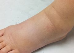
Many people with lymphedema or lipedema have no idea their condition has a name or that many other people suffer from the same thing. Although lymphedema and lipedema can’t be cured, proper management and resources can help patients cope. This article improves your grasp of these conditions, describes how to recognize and manage them, and explains how to support your patients.

To understand lymphedema and lipedema, first you need to understand how the lymphatic system functions. It makes lymph, then moves it from tissues to the bloodstream. It also plays a major role in the immune system, aiding immune defense. In addition, it helps maintain normal fluid balance by promoting fluid movement from the interstitial tissues back to the venous circulation. (See Lymphatic system: Four major functions.)

If the lymphatic system is impaired from a primary (hereditary or congenital) condition or a secondary problem, lymphedema can result. In this chronic, potentially progressive, and incurable condition, protein-rich fluid accumulates in the interstitial tissues.

Lymphedema basics

Lymphedema occurs in four stages.

Stage 0. During this stage (also called the subclinical or latency stage), transport capacity of the lymphatic system decreases but remains sufficient to manage normal lymphatic loads. Signs and symptomsaren’t evident and can be measured only by sensitive instruments, such as bioimpedance spectroscopy and optoelectronic volumetry. Without such instruments to quantify volume changes, diagnosis may rest on subjective complaints.

In this stage, limited functional reserve of the lymphatic system leads to a fragile balance between subnormal transport capacity and lymphatic loads. Added stress on the lymphatic system (as from extended heat or cold exposure, injury, or infection) may cause progression to stage 1.

Providing appropriate patient information and education, especially after surgery, can dramatically reduce the risk that lymphedema will progress to a more serious stage.

Stage 1. Considered the spontaneously reversible stage, stage 1 is marked by softtissue pliability without fibrotic changes. Pitting can be induced easily. In early stage 1, limb swelling may recede over – night. With proper management, the patient can expect the extremity to decrease to a normal size compared to that of the uninvolved limb. Otherwise, lymphedema is likely to progress to stage 2.

Stage 1 lymphedema may be hard to distinguish from edemas from other causes. Clinicians must rely on the patient history and monitor for swelling resolution with conventional management, such as compression and elevation, or note if swelling persists despite these standard interventions.

Stage 2. Sometimes called the spontaneously irreversible stage, stage 2 is identified mainly from tissue proliferation and subsequent fibrosis (called lymphostatic fibrosis). The fluid component can be removed spontaneously, but removal of the increased tissue proliferation (initially irreversible) takes more time. Tissue proliferation stems from long-standing accumulation of protein-rich fluid; over time, the tissue hardens and pitting is hard to induce. In many cases, swelling volume increases, exacerbating the already compromised local immune defense.

Consequently, infections (particularly cellulitis) are common; these, in turn, increase the volume of the affected area. Proper treatment can reduce volume.

With proper care (complete decongestive therapy [CDT]), lymphedema can stabilize during stage 2. But patients with chronic or recurrent infections are likely to progress to stage 3.

Stage 3. Also called lymphostatic elephantiasis, this stage is marked by further fluid volume increases and progression of tissue changes. Lymphostatic fibrosis becomes firmer and other skin alterations may occur, including papillomas, cysts, fistulas, hyperkeratosis, fungal infections, and ulcers. Pitting may be present. Natural skinfolds deepen (especially those of the dorsum of the wrist or ankle) and, in many cases, cellulitis recurs.

If lymphedema management starts during this stage, reduction can still occur. Even in extreme cases, with proper care and patient adherence to treatment, lymphostatic elephantiasis can be reduced so the leg is a normal or near-normal size.

Assessment and diagnosis

A thorough physical examination is the gold standard for diagnosing lymphedema. A complete patient history, body-systems review, inspection, and palpation can help determine if edema is lymphedema.

Clinically, the only test with proven reliability and validity in diagnosing lymphedema is the Stemmer sign. Fibrotic changes associated with lymphedema can lead to thickened skin over the proximal phalanges of the toes or fingers. If you can’t tent or pinch the skin on the involved extremity, lymphedema is present (a positive Stemmer sign). However, a negative finding (soft, pliable tissue) doesn’t rule out  lymphedema because the condition may be in an early stage, before tissue proliferation and fibrosis have set in.

Management

Although incurable, lymphedema can be managed successfully through CDT. This approach involves proper identification of lymphedema, manual lymph drainage, skin and nail care, patient education, compression, and exercise.

CDT has two phases:

Phase I, the intensive phase, continues until the extremity has decongested or reached a plateau. The clinician provides treatments and educates the patient about all aspects of CDT to prepare him or her for phase II. Phase I can last several weeks to several months depending on lymphedema severity.

Phase II, the maintenance phase, begins once the extremity has decongested or plateaued. This phase still focuses on CDT, but now the patient, not the clinician, is responsible for all care. The goal is to reduce limb size while enabling the patient to become self-sufficient in managing lymphedema. Although CDT can bring significant improvements in limb size, skin quality, and function, patients must remember that phase II continues lifelong. Be sure to provide education about ongoing self-management strategies.

Lipedema: The disease they call “fat”

Lipedema is a painful disorder of fat deposition. Pathologic deposition of fatty tissue (usually below the waist) leads to progressive leg enlargement. Like lymphedema, lipedema is incurable but manageable. Unless managed properly, lipedema can reduce mobility, interfere with activities of daily living, and lead to secondary lymphedema. (See Lipedema stages.)

Lipedema commonly is misdiagnosed as lymphedema. However, lymphedema involves protein-rich fluid, whereas lip edema is a genetically mediated fat disorder. Because lipedema resists diet and exercise, it can lead to psychosocial complications. Lipedema occurs almost exclusively in women; typically, onset occurs between puberty and age 30. One unpublished epidemiologic study puts lip edema incidence in females at 11%. Some patients have a combination of lipedema and lymphedema. (See Viewing lipolymphedema.)

Assessment and diagnosis

As with lymphedema, lipedema diagnosis rests on clinical presentation. Lipedema characteristics include bilateral and symmetrical involvement, absence of pitting (because lipedema isn’t a fluid disorder), soft and pliable skin, and filling of the retromalleolar sulcus (called the fat pad sign.)

Key signs and symptoms include:

• feeling of heaviness in the legs (aching dysesthesia)

• easy bruising

• sensitivity to touch (called “painful fat syndrome”)

• orthostatic edema

• oatmeal-like changes to skin texture.

Nearly half of lipedema patients are overweight or obese, but many appear of normal weight from the waist up. Essentially, the upper and lower extremities don’t match. The lower extremities typically show fatty deposits extending from the iliac crest to the ankles, sparing the feet. (See Lipedema patterns.)

Management

Lipedema is best  managed through weight control, as additional weight gain through adipose tissue tends to deposit in the legs. For patients with concomitant lymphedema (lipolymphedema), modified CDT helps reduce and manage lymphatic compromise. To address excess fat deposition, newer “wet” liposuction techniques have proven beneficial. These techniques gently detach adipose cells from the tissue, helping to preserve connective tissue and lymphatic vessels.

Know what to look for

In both lymphedema and lipedema, early identification and proper diagnosis are key. (See Differentiating lymphedema and lipedema.) A thorough history and physical exam will likely lead to an accurate diagnosis, if clinicians know what to look for. Proper diagnosis and treatment can prevent expensive and ineffective interventions, which can negatively affect both the patient’s condition and psychological well being.

Heather Hettrick is an associate professor at Nova Southeastern University, Department of Physical Therapy in Fort Lauderdale, Florida.

Selected references

Fat Disorders Research Society. Lipedema description.

Fife CE, Maus EA, Carter MJ. Lipedema: a frequently misdiagnosed and misunderstood fatty deposition syndrome. Adv Skin Wound Care. 2010;23(2):81-92

Herbst KL. Rare adipose disorders (RADS) masquerading as obesity. Acta Pharmacol Sin. 2012;33(2):155-72.

Lipedema Project.

National Lymphedema Network. Position papers.

Schmeller W, Hueppe M, Meier-Vollrath I. Tumescent liposuction in lipoedema yields good long-term results. Br J Dermatol. 2012;166(1):161-8.

Zuther J. A closer look at lipedema and the effects on the lymphatic system. December 13, 2012. lymphedemablog.com/2012/12/13/a-closer-look-at-lipedema-and-the-effects-on-the-lymphatic-system/

Zuther J. Stages of lymphedema. October 3, 2012.

Read More
1 2 3 4