Dragon blood may help wounds heal faster

synthetic peptide heal komodo dragon

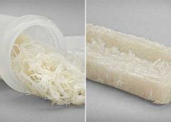
A new study has identified a peptide, derived from the Komodo dragon, called VK25, which can be synthesized and used as an antimicrobial peptide to promote wound healing.

The new research has identified (see below) a peptide found from the Komodo dragon (Varanus komodoensis), called VK25, which appears to be useful as a cationic antimicrobial peptide (CAMP). There is currently considerable interest in antimicrobial peptides in a world where antibiotic effectiveness is in decline. These peptides are potent, broad spectrum antibiotics which demonstrate potential as novel therapeutic agents. (more…)

Read More

Necrotizing Fasciitis: Pearls & Pitfalls

thigh fluid gas ct scan

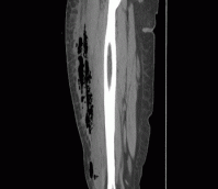
A 39-year-old woman presents to the ED with leg pain and fever. She initially noted redness and pain above her knee 2 weeks ago and was evaluated at an outside hospital. She completed a 10-day course of oral antibiotics for cellulitis. Over the last two days, she has had progressive leg swelling of her entire right thigh. The pain is now so severe that she is having difficulty walking. Her past medical history is negative for diabetes mellitus, chronic liver disease, or alcohol and IV drug use.

On exam, she is febrile to 102.7 F, heart rate is 96 bpm, and blood pressure is 112/65. She has a 12 cm area of faint erythema on her right thigh and tenderness to palpation of her entire right leg with diffuse edema. There is no ecchymosis or bullae formation. (more…)

Read More

Webinars

Winning the battle of skin tears in an aging population

"Skin tears" may sound like a relatively minor event, but in reality, these injuries can have a significant impact on the quality of patients' lives in the form of pain, infection, and limited mobility.

The incidence of skin tears has been reported to be as high as 1.5 million annually, and with an aging population, this number is likely to go higher.

In this webinar, experts will explain how nurses can use an evidence-based approach -- including following practice guidelines to assess the wound and select the proper dressing -- for managing skin tears and minimizing their negative effects.

[thrive_lead_lock id=’10942′][/thrive_lead_lock]

Skin Damage Associated with Moisture and Pressure

• Identify how wounds are classified according to wound depth and etiology

• Describe the etiology of pressure injury and incontinence- associated skin damage (IAD)

• Understand evidenced-based protocols of care for prevention and management of IAD and pressure injuries

• Recognize and describe NPUAP-EPUAP Pressure Injury Classification System

• Understand appropriate ConvaTec products that can be used for prevention and treatment of IAD and pressure injuries

[thrive_lead_lock id=’10974′][/thrive_lead_lock]

Read More

Pressure ulcer staging

By Nancy Morgan, RN, BSN, MBA, WOCN, WCC, CWCMS, DWC
Staging pressure ulcers can get tricky, especially when we’re dealing with a suspected deep-tissue injury (SDTI). The National Pressure Ulcer Advisory Panel defines an SDTI as a “purple or maroon localized area of discolored intact skin or blood-filled blister due to damage of underlying soft tissue from pressure and/or shear. The area may be preceded by tissue that is painful, firm, mushy, boggy, warmer, or cooler as compared to adjacent tissue… Deep tissue injury may be difficult to detect in individuals with dark skin tones. Evolution may include a thin blister over a dark wound bed. The wound may further evolve and become covered by thin eschar. Evolution may be rapid, exposing additional layers of tissue even with optimal treatment.” (more…)

Read More

How are you differentiating the “big three”?

BY: NANCY MORGAN, RN, BSN, MBA, WOCN, WCC, CWCMS, DWC
Lower extremity ulcers are often referred as the “big three”—arterial ulcers, venous ulcers, and diabetic foot ulcers. Are you able to properly identify them based on their characteristics? Sometimes, it’s a challenge to differentiate them.

Arterial ulcers tend occur the tips of toes, over phalangeal heads, around the lateral malleolus, on the middle portion of the tibia, and on areas subject to trauma. These ulcers are deep, pale, and often necrotic, with minimal granulation tissue. Surrounding skin commonly is pale, cool, thin, and hairless; toenails tend to be thick. Arterial ulcers tend to be dry with minimal drainage, and often are associated with significant pain. The patient usually has diminished or absent pulses. (more…)

Read More

If All You Have is a Hammer, What Happens When You Run Out of Nails?

hammer

by Dr. Michael Miller

Over the years of making house calls for wound care, I found that there was a real need for home based mental health and behavioral care, palliative care, podiatry and lots of other things. We cater to those who are home bound based on the classic definition involving the word “Taxing”. One of the more prevalent problems affecting all patients involves the nebulous but ubiquitous, nerve jangling, aptly named, “5th Vital Sign”, namely pain. As a part of my medical group, we have created a program that provides pain management not just to the home bound but all those whose lives and lifestyles are affected adversely by it. The program is a monument to government bureaucracy involving multiple layers of paperwork, mental health evaluations, testing of bodily fluids for both illegal and legal substances and then, the actual evaluation of the patient commences. After all hurdles are vetted and then jumped, then and only then does a prescription for the appropriate nostrum leave the pad. In wound care, we treat based on the etiology, the location, the related factors, the amounts of drainage, the surrounding tissues and so on, ad nauseum. Not surprisingly, in pain management, the scenario is much different. In wound care the mantra of the dabbler is see the hole, fill the hole. In pain management, the goal is to minimize pain to maximize functionality but the overriding questions are how this is accomplished. (more…)

Read More

Don’t Kid Yourself, Amputation Is Unquestionably A Failure

amputation is a failure

by Dr. Michael Miller

I recently saw an ad for a pending lecture at a national conference that piqued my interest much like “deflate-gate”.  The title of this lecture horrifically touted that Amputation need not be considered failure.  As a full time wound care doc, I work to identify those conditions that place patients at risk of all consequences both limited and catastrophic.  We use the catchy title of “Limb Preservation”.  We start the process by engaging in the unusual behavior of making definitive diagnoses, then systematically address them in as comprehensive manner as possible.  I am proud to tell you that while there are occasions in which a terminally damaged digit is lost,  that we have rarely sacrificed the greater part of a foot and more, have had only 3 lower extremity amputations in the last 5 years on patients who’s care remained exclusively with us.  Of course, when a patient for whom we have created and implemented a “Limb Pres” care plan is taken out of our system (usually via a hospitalization for a reason other then the lower extremity problem), the facility forces that be unfortunately but infrequently demonstrate their inadequacy and paranoia by gang-harangueing the patient and family.  They are lambasted with lurid tales of the condition marching up the leg engulfing the foot, knee, torso, and brains much like a flesh-eating PacMan.   The patient’s confidence now neutered has little chance against this persistent onslaught of inadequacy and so, much like the Queen song, “Another One Bites The Dust”. (more…)

Read More

eBooks

Creating an Ideal Microenvironment for Wound Cleansing

[Sponsored by Angelini Pharma, Inc.]

With so much focus on dressing choices, it’s easy to forget the importance of wound cleansing. Wound cleansing can help achieve the goals of wound bed preparation by removing microorganisms, biological and environmental debris to create an environment beneficial to healing as well as facilitating wound assessment by allowing clear visualization of the wound.

Preventing Infections in Patients with Wounds eBook

[Sponsored by Angelini Pharma, Inc.]

Proper wound care is essential to preventing infections for patients in all practice settings, and healthcare providers should stay informed about the most current and effective treatments out there.

Recognizing factors that increase patients’ susceptibility to infection allows providers to identify risks and take measures to prevent infection from occurring or worsening.

eBook: Treatment of Hard-to-Heal Wounds with Collagen-Based Dressings

[Sponsored by Angelini Pharma, Inc.]

Receive a free BioPad Sample and a free eBook with more details.

BioPad Wound Dressing with Collagen: BioPad, is a 100% equine Type-1 collagen primary wound dressing used to treat hard to heal wounds.

The BioPad collagen dressing has the highest collagen content on the market up to 5 times the amount of collagen. 

BioPad™ is the perfect dressing for wounds.

eBook: Caring for Wounds eBook Series: Pressure Injuries

[Sponsored by Angelini Pharma, Inc.]

Learn how your healthcare team can provide better patient care.

Patient care teams rely on the wound care nurse alone to implement a pressure ulcer prevention program; however, a successful program requires involvement from the entire care team and is a 24/7 endeavor.

eBook: Skin Damage Associated with Moisture and Pressure

[Sponsored by Convatec]

Tips on how to differentiate and goals for protection and management.

* Identify how wounds are classified according to wound depth and etiology.
* Describe the etiology of a pressure injury (PI) and incontinence-associated skin damage (IAD).
* Discuss evidence-based protocols of care of prevention and management if IAD and PIs.
* Describe the NPUAP-EPUAP Pressure Injury Classification System.
* Identify appropriate products that can be used for preventioin and treatment of IAD and PIs.

eBook: Needlestick Risks: Defense and Rescue Strategies for Nurses

[This e-book has been developed through an educational grant from CM&F Group]

Learn more about: 
A Continuing Risk for Healthcare Workers, Sharps Injuries: Facts and Figures, Proactive Steps for Yourself and Your Colleagues, A Preventable Injury, A Downloadable Workbook from the CDC, The Case for Coverage, If You are Exposed.

Needlesticks and other sharps-related exposures to bloodborne pathogens (including HIV, hepatitis B virus, and hepatitis C virus) continue to pose a significant occupational risk for healthcare workers


Safe Biopsies eBook: Protect yourself and your patients.

[This e-book is brought to you by BiopSafe]

Safe biopsy handling
One of the most common problems in connection with biopsy handling is the risk of being exposed to formalin either through touch or inhalation. A risk that doctors, veterinarians, laboratory technicians and nurses are exposed to every day.

With BiopSafe the problem is finally solved.

receive a free BiopSafe Sample and a free eBook PDF with more information and details.

 
Read More

Breaking silos: Effective wound healing means treatment across the continuum

Around 6.5 million patients in the U.S. suffer from chronic wounds, such as pressure injuries or ulcers. Treatment costs $25 billion each year, representing a sizable and growing problem. Despite the wide impact of chronic wounds, it’s rare to see specialized, effective wound care delivered across the care continuum.

A chronic non-healing wound is a surrogate marker for illness. These patients require holistic management of their co-morbidities and continuity across care settings.

Despite this, a great deal of emphasis has been placed on treating wounds as singular events, managed topically with expensive dressings and support surfaces. This is only a small part of wound healing.

As a physician focused solely on wound care, I have learned that we must shift the focus from simply treating the wound to treating the wounded patient. The impact in the post-acute care setting in particular is worthy of evaluation and discussion, as up to 29% of patients in long-term care facilities will experience a pressure ulcer, posing serious legal, financial, and staffing implications.

For those providers working outside long-term care, there is little understanding of challenges facing LTC providers. Acute providers do not often ask, for example, how are my LTC partners reimbursed? How are they staffed? What are the requirements and regulatory pressures they face? Asking these questions would facilitate a more productive dialogue with a focus on collaborative prevention, rather than waiting until a chronic wound occurs in the LTC setting.

Creating an integrated wound care community

To address the needs of the present and growing population of patients with chronic wounds, Healogics developed an integrated wound care community model, to coordinate the wound healing process across all care settings. The program utilizes Healogics Specialty Physicians, a subspecialty group of physicians and providers with extensive training solely focused on wound care.

HSPs provide expert inpatient consultation and ensure safe transition of patients out of the hospital into the appropriate care setting. Because HSPs see the patient regardless of post-discharge venue, patients receive the same quality of care whether they are going home, to a skilled-nursing, assisted living, or LTC setting. Because chronic wounds are surrogate markers for illness, we have realized it’s essential to have an integrated, multi-setting, and multi-disciplinary process to treat the patient and their co-morbidities.

Data collected at a pilot IWCC site in the Midwestern U.S. from 2014 to 2016 revealed very positive trends for chronic wound patients. In the acute care setting, the average length of stay decreased from 9.41 days to 5.64 days, and total cost of care per patient was reduced from $10,670 to $7,248.

We’re excited by these promising results, which were revealed at the American College of Wound Healing and Tissue Repair Conference last December. We look forward to refining and expanding the model by helping our partners in acute and LTC settings standardize their practices, use evidence-based clinical guidelines, mobilize technologies and processes, and pay critical attention to patient safety and value-based outcomes.

When it comes to wound healing, no venue of care should operate alone—an integrated solution that creates continuity for the patient is critical. There are four things LTC facilities can do to break down the silos:

Read more at McKnight’s

Read More

Herpes zoster: Understanding the disease, its treatment, and prevention

Herpes zoster: Understanding the disease, its treatment, and prevention

Herpes zoster (HZ, also called shingles) is a painful condition that produces a maculopapular and vesicular rash. Usually, the rash appears along a single dermatome (band) around one side of the body or face.

In most cases, pain, tingling, burning, or itching occurs a few days before the rash. Next, blisters form, scabbing over in 7 to 10 days. In rare cases, the rash is widespread, resembling varicella zoster (VZ, or chickenpox) rash. Pain can range from mild to severe and may be dull, burning, or gnawing. It may last weeks, months, or even years after the blisters heal. Shingles on the face may impair vision or hearing. (more…)

Read More

Lymphedema and lipedema: What every wound care clinician should know

Imagine you have a health condition that affects your life every day. Then imagine being told nothing can be done about it; you’ll just have to live with it. Or worse yet, your physician tells you the problem is “you’re just fat.”

Many people with lymphedema or lipedema have no idea their condition has a name or that many other people suffer from the same thing. Although lymphedema and lipedema can’t be cured, proper management and resources can help patients cope. This article improves your grasp of these conditions, describes how to recognize and manage them, and explains how to support your patients.

To understand lymphedema and lipedema, first you need to understand how the lymphatic system functions. It makes lymph, then moves it from tissues to the bloodstream. It also plays a major role in the immune system, aiding immune defense. In addition, it helps maintain normal fluid balance by promoting fluid movement from the interstitial tissues back to the venous circulation. (See Lymphatic system: Four major functions.)

If the lymphatic system is impaired from a primary (hereditary or congenital) condition or a secondary problem, lymphedema can result. In this chronic, potentially progressive, and incurable condition, protein-rich fluid accumulates in the interstitial tissues.

Lymphedema basics

Lymphedema occurs in four stages.

Stage 0. During this stage (also called the subclinical or latency stage), transport capacity of the lymphatic system decreases but remains sufficient to manage normal lymphatic loads. Signs and symptomsaren’t evident and can be measured only by sensitive instruments, such as bioimpedance spectroscopy and optoelectronic volumetry. Without such instruments to quantify volume changes, diagnosis may rest on subjective complaints.

In this stage, limited functional reserve of the lymphatic system leads to a fragile balance between subnormal transport capacity and lymphatic loads. Added stress on the lymphatic system (as from extended heat or cold exposure, injury, or infection) may cause progression to stage 1.

Providing appropriate patient information and education, especially after surgery, can dramatically reduce the risk that lymphedema will progress to a more serious stage.

Stage 1. Considered the spontaneously reversible stage, stage 1 is marked by softtissue pliability without fibrotic changes. Pitting can be induced easily. In early stage 1, limb swelling may recede over – night. With proper management, the patient can expect the extremity to decrease to a normal size compared to that of the uninvolved limb. Otherwise, lymphedema is likely to progress to stage 2.

Stage 1 lymphedema may be hard to distinguish from edemas from other causes. Clinicians must rely on the patient history and monitor for swelling resolution with conventional management, such as compression and elevation, or note if swelling persists despite these standard interventions.

Stage 2. Sometimes called the spontaneously irreversible stage, stage 2 is identified mainly from tissue proliferation and subsequent fibrosis (called lymphostatic fibrosis). The fluid component can be removed spontaneously, but removal of the increased tissue proliferation (initially irreversible) takes more time. Tissue proliferation stems from long-standing accumulation of protein-rich fluid; over time, the tissue hardens and pitting is hard to induce. In many cases, swelling volume increases, exacerbating the already compromised local immune defense.

Consequently, infections (particularly cellulitis) are common; these, in turn, increase the volume of the affected area. Proper treatment can reduce volume.

With proper care (complete decongestive therapy [CDT]), lymphedema can stabilize during stage 2. But patients with chronic or recurrent infections are likely to progress to stage 3.

Stage 3. Also called lymphostatic elephantiasis, this stage is marked by further fluid volume increases and progression of tissue changes. Lymphostatic fibrosis becomes firmer and other skin alterations may occur, including papillomas, cysts, fistulas, hyperkeratosis, fungal infections, and ulcers. Pitting may be present. Natural skinfolds deepen (especially those of the dorsum of the wrist or ankle) and, in many cases, cellulitis recurs.

If lymphedema management starts during this stage, reduction can still occur. Even in extreme cases, with proper care and patient adherence to treatment, lymphostatic elephantiasis can be reduced so the leg is a normal or near-normal size.

Assessment and diagnosis

A thorough physical examination is the gold standard for diagnosing lymphedema. A complete patient history, body-systems review, inspection, and palpation can help determine if edema is lymphedema.

Clinically, the only test with proven reliability and validity in diagnosing lymphedema is the Stemmer sign. Fibrotic changes associated with lymphedema can lead to thickened skin over the proximal phalanges of the toes or fingers. If you can’t tent or pinch the skin on the involved extremity, lymphedema is present (a positive Stemmer sign). However, a negative finding (soft, pliable tissue) doesn’t rule out  lymphedema because the condition may be in an early stage, before tissue proliferation and fibrosis have set in.

Management

Although incurable, lymphedema can be managed successfully through CDT. This approach involves proper identification of lymphedema, manual lymph drainage, skin and nail care, patient education, compression, and exercise.

CDT has two phases:

Phase I, the intensive phase, continues until the extremity has decongested or reached a plateau. The clinician provides treatments and educates the patient about all aspects of CDT to prepare him or her for phase II. Phase I can last several weeks to several months depending on lymphedema severity.

Phase II, the maintenance phase, begins once the extremity has decongested or plateaued. This phase still focuses on CDT, but now the patient, not the clinician, is responsible for all care. The goal is to reduce limb size while enabling the patient to become self-sufficient in managing lymphedema. Although CDT can bring significant improvements in limb size, skin quality, and function, patients must remember that phase II continues lifelong. Be sure to provide education about ongoing self-management strategies.

Lipedema: The disease they call “fat”

Lipedema is a painful disorder of fat deposition. Pathologic deposition of fatty tissue (usually below the waist) leads to progressive leg enlargement. Like lymphedema, lipedema is incurable but manageable. Unless managed properly, lipedema can reduce mobility, interfere with activities of daily living, and lead to secondary lymphedema. (See Lipedema stages.)

Lipedema commonly is misdiagnosed as lymphedema. However, lymphedema involves protein-rich fluid, whereas lip edema is a genetically mediated fat disorder. Because lipedema resists diet and exercise, it can lead to psychosocial complications. Lipedema occurs almost exclusively in women; typically, onset occurs between puberty and age 30. One unpublished epidemiologic study puts lip edema incidence in females at 11%. Some patients have a combination of lipedema and lymphedema. (See Viewing lipolymphedema.)

Assessment and diagnosis

As with lymphedema, lipedema diagnosis rests on clinical presentation. Lipedema characteristics include bilateral and symmetrical involvement, absence of pitting (because lipedema isn’t a fluid disorder), soft and pliable skin, and filling of the retromalleolar sulcus (called the fat pad sign.)

Key signs and symptoms include:

• feeling of heaviness in the legs (aching dysesthesia)

• easy bruising

• sensitivity to touch (called “painful fat syndrome”)

• orthostatic edema

• oatmeal-like changes to skin texture.

Nearly half of lipedema patients are overweight or obese, but many appear of normal weight from the waist up. Essentially, the upper and lower extremities don’t match. The lower extremities typically show fatty deposits extending from the iliac crest to the ankles, sparing the feet. (See Lipedema patterns.)

Management

Lipedema is best  managed through weight control, as additional weight gain through adipose tissue tends to deposit in the legs. For patients with concomitant lymphedema (lipolymphedema), modified CDT helps reduce and manage lymphatic compromise. To address excess fat deposition, newer “wet” liposuction techniques have proven beneficial. These techniques gently detach adipose cells from the tissue, helping to preserve connective tissue and lymphatic vessels.

Know what to look for

In both lymphedema and lipedema, early identification and proper diagnosis are key. (See Differentiating lymphedema and lipedema.) A thorough history and physical exam will likely lead to an accurate diagnosis, if clinicians know what to look for. Proper diagnosis and treatment can prevent expensive and ineffective interventions, which can negatively affect both the patient’s condition and psychological well being.

Heather Hettrick is an associate professor at Nova Southeastern University, Department of Physical Therapy in Fort Lauderdale, Florida.

Selected references

Fat Disorders Research Society. Lipedema description.

Fife CE, Maus EA, Carter MJ. Lipedema: a frequently misdiagnosed and misunderstood fatty deposition syndrome. Adv Skin Wound Care. 2010;23(2):81-92

Herbst KL. Rare adipose disorders (RADS) masquerading as obesity. Acta Pharmacol Sin. 2012;33(2):155-72.

Lipedema Project.

National Lymphedema Network. Position papers.

Schmeller W, Hueppe M, Meier-Vollrath I. Tumescent liposuction in lipoedema yields good long-term results. Br J Dermatol. 2012;166(1):161-8.

Zuther J. A closer look at lipedema and the effects on the lymphatic system. December 13, 2012. lymphedemablog.com/2012/12/13/a-closer-look-at-lipedema-and-the-effects-on-the-lymphatic-system/

Zuther J. Stages of lymphedema. October 3, 2012.

Read More
1 2 3 4 5 9