BY: NANCY MORGAN, RN, BSN, MBA, WOCN, WCC, CWCMS, DWC
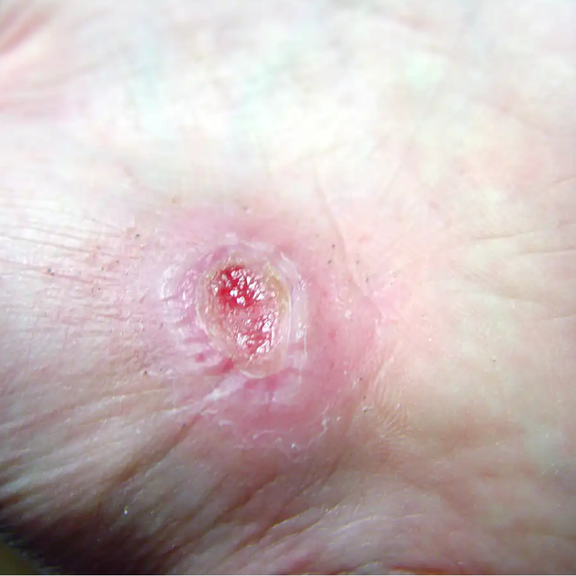
Lower extremity ulcers are often referred as the “big three”—arterial ulcers, venous ulcers, and diabetic foot ulcers. Are you able to properly identify them based on their characteristics? Sometimes, it’s a challenge to differentiate them.
Arterial ulcers tend occur the tips of toes, over phalangeal heads, around the lateral malleolus, on the middle portion of the tibia, and on areas subject to trauma. These ulcers are deep, pale, and often necrotic, with minimal granulation tissue. Surrounding skin commonly is pale, cool, thin, and hairless; toenails tend to be thick. Arterial ulcers tend to be dry with minimal drainage, and often are associated with significant pain. The patient usually has diminished or absent pulses.
Venous ulcers are located on the medial lower leg, medial malleolus, and superior to the medial malleolus. You rarely see them on the foot or above the knee. They have irregular wound margins and tend to be shallow and ruddy red, although slough may be present. Venous ulcers tend to have moderate to large drainage amounts. Although they don’t usually cause a lot of pain, patients may complain of “achy” legs. Surrounding skin is scaly and weepy, possibly with hemosiderin staining and edema. The patient usually has palpable pulses.
Diabetic foot ulcers arise on the plantar aspect of the foot, over metatarsal heads, and under the heel. They have even wound margins and often are deep ulcers with red or pale granular wound beds. Slough is common. Surrounding tissue is often a callus, and cellulitis is common. A low to moderate amount of drainage is present, and foot deformities are common. The patient typically has diminished or absent sensation in the foot. Due to vessel calcifications, we don’t rely on the ankle brachial index (ABI) for these patients. Instead, we use the toe brachial pressure index (TBPI).
Even though arterial, venous, and diabetic ulcers have specific characteristics, not all individual wounds follow the rules. We may see mixed ulcers types with components of both arterial and venous assessment findings. Diabetic foot ulcers may look arterial, especially when the toe is necrotic.
Do you rely just on your visual assessment of these types of ulcers, or do you use diagnostic testing to differentiate them? What diagnostics are you using? Do all of your patients with lower extremity ulcers get ABIs? Are you obtaining TBPIs on your diabetic patients?
DISCLAIMER: All clinical recommendations are intended to assist with determining the appropriate wound therapy for the patient. Responsibility for final decisions and actions related to care of specific patients shall remain the obligation of the institution, its staff, and the patients’ attending physicians. Nothing in this information shall be deemed to constitute the providing of medical care or the diagnosis of any medical condition. Individuals should contact their healthcare providers for medical-related information.

