Qualified healthcare professionals (QHPs), such as physicians, podiatrists, physician assistants, nurse practitioners, and clinical nurse specialists, are taught to diagnose the reasons that chronic wounds aren’t healing and to create plans of care for aggressively managing the wound until it heals. Wound care professionals—nurses and therapists—are taught to implement those plans of care. All of these highly skilled wound care professionals know how to manage chronic wounds from identification through healing. (more…)
As a senior quality improvement specialist with IPRO, the Quality Improvement Organization for New York State over the past 11 years, I’ve been tasked with helping skilled nursing facilities (SNFs) embrace the process of continuous quality improvement. A necessary component of this effort has been to collect, understand, and analyze timely and accurate data. This article discusses a free tool I developed to help SNFs track their data related to pressure ulcers and focus their quality improvement efforts for the greatest impact. (more…)
Each issue, Apple Bites brings you a tool you can apply in your daily practice.
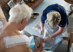
Measurement of wounds is an important component of wound assessment and provides baseline measurements, enables monitoring of healing rates, and helps distinguish among wounds that are static, deteriorating, or improving. All alterations in skin integrity, including those caused by ulcers, venous ulcers, arterial ulcers, neuropathic ulcers, incision lines, grafts, donor sites, abscesses, and rashes should be measured when they’re discovered and at intervals thereafter, based on institutional policy. (more…)
Pressure-ulcer prevention and management guidelines recommend support-surface therapy to help prevent and treat pressure ulcers. Support surfaces include pads, mattresses, and cushions that redistribute pressure. Full cushions and cushion pads are considered therapeutic support surfaces if used to redistribute a patient’s pressure in a chair or wheelchair.
The National Pressure Ulcer Advisory Panel (NPUAP) defines support surfaces as “specialized devices for pressure redistribution designed for the management of tissue loads, microclimate, and/or other therapeutic functions.” These surfaces address the mechanical forces associated with skin and tissue injury, such as pressure, shear, friction, and excess moisture and heat. (See Clearing up the confusion.)
Incidence density best measure of pressure-ulcer prevention program According to the National Pressure Ulcer Advisory Panel (NPUAP), incidence density is the best quality measure of pressure-ulcer prevention programs. Pressure-ulcer incidence density is calculated by dividing the number of inpatients who develop a new pressure ulcer by 1,000 patient days. Using the larger denominator of patient days allows fair comparisons between institutions…
This issue’s resources include patient tools and new guidelines. Improving patient safety Research suggests that adverse events affect patients with limited English proficiency (LEP) more frequently, are commonly caused by communication problems, and are more likely to result in serious harm compared to adverse events affecting English-speaking patients. Your hospital can take steps to reduce risks of adverse events for…
By Debra Rose Wilson, PhD, MSN, RN, IBCLC, AHN-BC, and Dana Marie Dillard, MS, HSMI Like many clinicians, you may experience stress frequently, both on and off the job. Chronic stress can alter your equilibrium (homeostasis), activating physiologic reactive pathways that cause your body to shift its priorities. Physiologic effects of stress may include: slowed digestion delay in reproductive and…
By Pamela Anderson, MS, RN, APN-BC, CCRN, and Terri Townsend, MA, RN, CCRN-CMC, CVRN-BC Jan Smith, age 59, is admitted to the coronary intensive care unit with an acute inferior myocardial infarction (MI). Recently diagnosed with hypertension and hyperlipidemia, she smokes a pack and a half of cigarettes daily. She reports she has always been healthy and can’t believe she…
By: Donna Sardina, RN, MHA, WCC, CWCMS, DWC, OMS Years ago, when I first started out in the wound care specialty, the only way to learn about new products and what was going on in the field was to “go to conference” (wound care conference). All year long, planning and excitement continued to build for our big trip. Not going…
By Cheryl Ericson, MS, RN, CCDS, CDIP Providers are often surprised at how pages upon pages of documentation in a patient’s health record can result in few reportable diagnosis and/or procedure codes, which often fail to capture the complexity of the patient’s condition. However, providers need to be aware of the implications of coding. As healthcare data become increasingly digital…
By Catherine R. Ratliff, PhD, APRN-BC, CWOCN, CFCN It’s estimated that about 70% of the 1 million ostomates in the United States and Canada will experience or have experienced stomal or peristomal complications. Peristomal complications are more common, although stomal complications (for example, retraction, stenosis, and mucocutaneous separation) can often contribute to peristomal problems by making it difficult to obtain…
By Nancy Morgan, RN, BSN, MBA, WOC, WCC, DWC, OMS Each issue, Apple Bites brings you a tool you can apply in your daily practice. The crusting procedure produces a dry surface and absorbs moisture from broken skin through an artificial scab that’s created by using skin barrier powder (stoma powder) and liquid polymer skin barrier. The crusting procedure is…
By Rosalyn S. Jordan, BSN, RN, MSc, CWOCN, WCC, and Sandra Phipps, BSN, RN, MBA, WCC Pressure-ulcer prevention and management guidelines recommend support-surface therapy to help prevent and treat pressure ulcers. Support surfaces include pads, mattresses, and cushions that redistribute pressure. Full cushions and cushion pads are considered therapeutic support surfaces if used to redistribute a patient’s pressure in a…
By Jeri Lundgren, BSN, RN, PHN, CWS, CWCN Prevention of pressure ulcers and skin breakdown begins with a comprehensive risk assessment. Most providers use a skin risk assessment tool, such as the Braden or Norton scale. While these tools have been validated to predict pressure ulcer development, their use alone isn’t considered a comprehensive assessment, and frequently the individual risk…
By Carrie Carls, BSN, RN, CWOCN, CHRN, and Sherry Clayton, RHIA
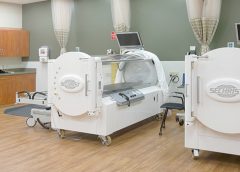
In an atmosphere of changing reimbursement, it’s important to understand indications and utilization guidelines for healthcare services. Otherwise, facilities won’t receive appropriate reimbursement for provided services. This article focuses on Medicare reimbursement for hyperbaric oxygen therapy (HBOT). (See What is hyperbaric oxygen therapy?)
By Jennifer Zinn, MSN, RN, CNS-BC, CNOR, and Vangela Swofford, BSN, RN, ASQ-CSSBB
For surgical patients, operative wound classification is crucial in predicting postoperative surgical site infections (SSIs) and associated risks. Information about a patient’s wound typically is collected by circulating registered nurses (RNs) and documented at the end of every surgical procedure. (more…)
A pressure ulcer that a patient acquires in your facility or a patient’s existing pressure ulcer that worsens puts your organization at risk for regulatory citations as well as litigation. Unless you can prove the pressure ulcer was unavoidable, you could find yourself burdened with citations or fines, or could even end up in court. (more…)
Critical limb ischemia may not increase mortality risk in patients with diabetes
Diabetic patients with critical limb ischemia (CLI) who are assessed quickly and treated aggressively do not have an increased risk of long-term cardiac mortality, according to a study in Diabetes Care.(more…)
What an honor it is to be the wound care “superhero”—the guru, the healer, the go-to person. Unfortunately, this honor may be accompanied by wound care overload—too much to do in too little time.
Once someone is crowned the superhero specialist, others may try to transfer every aspect of wound and skin care to that person—all treatment plans, assessments, documentation, prevention, education, and accountability. Superheroes don’t cry, so they don’t complain about the workload. Yet, the overload must be controlled. (more…)
By Rosalyn Jordan, RN, BSN, MSc, CWOCN, WCC, and Marci Christian, BBE
Any patient with a fecal or urinary ostomy may experience complications on the skin surface around the stoma. These complications may occur lifelong, although they’re more common during the first 5 years after the initial ostomy surgery. Causative factors include infection, trauma, certain diseases, and chemical irritation; most of these problems stem from the pouching system or pouch leakage.
Peristomal skin complications can cause a wide range of signs and symptoms, from skin discoloration to polyp-like growths, from erythema to full-thickness wounds. They can lead to discomfort, pain, poor self-image, social isolation, and impaired quality of life, not to mention additional care costs.
Incidence and types of these complications are hard to compare or contrast across multiple patients. Until recently, no standardized assessment or documentation tools were available to characterize or define complications. For this reason, reported rates ranged widely, from 10% to 70%. And because no designated common language or categories related to peristomal skin complications existed, documentation was inconsistent.
In the late 2000s, a group of nurses experienced in caring for ostomy patients worked with the World Council of Enterostomal Therapists to develop a resource called the Ostomy Skin Tool, which clinicians can use to categorize and describe peristomal skin complications in a consistent, objective manner. The tool also provides a common language for documentation.
The Ostomy Skin Tool has three major assessment domains—discoloration (D), erosion/ulceration (E), and tissue overgrowth (T), known collectively as DET. The DET combined rating ranges from normal, rated 0, to the worst condition possible, rated 15. Mild DET complications are documented as less than 4, moderate as less than 7, and severe as 8 or higher. (See Using the Ostomy Skin Tool by clicking the PDF icon above.)
The tool describes four categories of peristomal complications:
• chemical irritation
• mechanical trauma
• disease-related complications
• infection-related complications.
Chemical irritation
Chemical irritation can stem from irritants (as in contact dermatitis) or allergic reactions (allergic dermatitis). The most likely cause of chemical dermatitis is effluent leakage (feces or urine) from the colostomy, ileostomy, or urostomy, in which effluent comes in contact with peristomal skin. Other potential causes include contact with soap, certain adhesives, and adhesive removers.
The major treatment of chemical irritation is identification and removal of the offending agent, followed by patient and caregiver education on the new pouching procedure the patient must use. Follow-up assessment also is recommended. In a 2010 study that followed 89 patients for 1 year after ostomy surgery, about 50% of subjects experienced peristomal skin complications, most of them from pouch leakage. Another investigator estimated that 85% of ostomy patients experience pouch leakage at some time during their lives. Pouch leakage usually occurs when stool is extremely liquid (for instance, ileostomy effluent). Other causes of pouch leakage include wearing a pouch more than half full of effluent and abdominal contours that aren’t level. Besides changes in the pouching system, treatment may entail adding products to the pouching system or removing certain agents.
Some patients experience allergic dermatitis in reaction to products used in the pouching system (such as skin barriers, belts, pouch closures, or adhesives). However, allergic dermatitis is rare. One 2010 study suggested allergic reactions to these products occur in only about 0.6% of patients with peristomal skin irritation. Most major ostomy product manufacturers provide a patch test on request to help identify allergic conditions. Once the offending product is discontinued, allergic dermatitis should resolve rapidly.
Mechanical trauma
Mechanical trauma usually results from either the pouching system itself or its removal. It also may result from harsh or multiple skin-barrier removals, pressure from convex rings or pouches, and abrasive cleansing techniques. Some researchers believe the stronger the adhesive barrier and the more often a pouch is changed, the greater the risk of epidermal damage.
Mechanical trauma may present as a partial-thickness ulcer caused by pressure, shear, friction, tearing, or skin stripping. Patients with fragile skin are susceptible to mechanical trauma, so less aggressive pouching systems may be preferred for them. Of course, if the pouching system is changed, the patient or caregiver needs to learn about the new system.
Disease-related complications
Disease-related peristomal complications may be linked to preexisting skin conditions, such as psoriasis, eczema (atopic dermatitis), or seborrheic dermatitis. Hyperplasia also may occur. This overgrowth of cells, which may appear as gray or reddish brown pseudoverrucous lesions, usually is linked to urinary ostomies, although it can occur with fecal ostomies as well. Vinegar soaks are the recommended treatment, in addition to a change in the pouching system and corresponding patient education.
Occasionally, other disease-related complications occur, including primary adenocarcinoma of the peristomal skin and peristomal pyoderma gangrenosum, a painful and problematic condition that presents as peristomal ulcers. Ulcer borders are well-defined with a bluish purple coloration at the edges. Infection must be ruled out, as this condition usually is linked to an autoimmune condition. Treatment includes pain management and, in most cases, a topical corticosteroid. Crohn’s disease also may manifest as a peristomal skin ulcer.
Infection-related complications
Infection-related complications may be bacterial or fungal. Two common peristomal skin infections are folliculitis and Candida fungal infections. An infection of the hair follicle that causes pustules, folliculitis usually stems from traumatic hair pulling in the peristomal area during pouch removal. It may warrant a prescribed antibiotic, along with patient teaching regarding proper hair removal using an electric razor.
Candida infections may arise because peristomal skin provides a warm, dark, moist environment that promotes fungal growth. These infections appear as erythema with pustules or papules and satellite lesions. Treatment usually involves antifungal powder and use of the crusting technique to secure the pouching system. (See Using the crusting technique by clicking the PDF icon above.)
Management
Many complications are well advanced by the time patients seek assistance, perhaps because they don’t understand the significance of their symptoms and think they can manage the problem themselves. In some cases, they don’t know where to turn for assistance. Commonly, the complication progresses to the point where the patient goes to the emergency department or (particularly during the immediate postoperative period) needs to be readmitted for treatment. The best way to manage peristomal skin complications is to prevent them in the first place. (See Preventing peristomal skin complications by clicking the PDF icon above.)
Patient education
Over the past 20 years, hospital stays for ostomy surgery patients have decreased from about 2 weeks to less than 5 days. Reduced stays decrease the time available for caregivers to teach patients and family members how to empty and change the pouch. They need alternative education covering (among other topics) how to recognize peristomal skin complications and when to seek help. Not only do these complications require vigilant self-observation, but many patients don’t understand their implications or how rapidly they can worsen. In some cases, the first symptoms are itching and redness under the skin barrier. Fortunately, some patients may know or remember that itching, burning, stinging, reddened, or weeping peristomal skin requires professional attention. They can avoid serious complications by seeking assistance early, such as right after noticing pouch leakage.
Early treatment can reduce the cost of treatment. In a 2012 study, researchers estimated care costs related to peristomal skin complications for a 7-week treatment period, using the Ostomy Skin Tool as a reference. Severe complications (those with a DET score above 8) cost six times more to treat than mild cases (those with a DET score below 4) and 4.5 times more than moderate cases.
Along with early intervention by a trained ostomy care specialist, self-assessment by ostomy patients promotes a better quality of life, reduces pain, and may decrease care costs. Clinicians’ use of the Ostomy Skin Tool to assess and document peristomal skin complications promotes more reliable, objective, comparable assessment data for reporting.
Selected references
Al-Niaimi F, Lyon CC. Primary adenocarcinoma in peristomal skin: a case study. Ostomy Wound Manage. 2010;56(1):45-7.
Burch J. Management of stoma complications. Nurs Times. 2011;107(45):17-8, 20.
Jemec GB, Martins L, Claessens I, et al. Assessing peristomal skin changes in ostomy patients: validation of the Ostomy Skin Tool. Br J Dermatol. 2011; 164;330-5.
Jones T, Springfield T, Brudwick M, Ladd A. Fecal ostomies: practical management for the home health clinician. Home Healthc Nurse. 2011;29(5):306-17.
Martins L, Samai O, Fernandez A, et al. Maintaining healthy skin around an ostomy: peristomal skin disorders and self-assessment. Gastrointest Nurs. 2011;
9(2):9-13.
Martins L, Tavernelli K, Serrano JLC. Introducing a peristomal skin assessment tool: The Ostomy Skin Tool. World Council Enterostomal Therapists J. 2008;28(2):3-13.
Meisner S, Lehur P, Moran B, et al. Peristomal skin complications are common, expensive, and difficult to manage: a population based cost modeling study. PLoS One. 2012;7(5):e37813.
Omura Y, Yamabe M, Anazawa S. Peristomal skin disorders in patients with intestinal and urinary ostomies: influence of adhesive forces of various hydrocolloid wafer skin barriers. J Wound Ostomy Continence Nurs. 2010;37(3):289-98.
Ratliff CR. Early peristomal skin complications reported by WOC nurses. J Wound Ostomy Continence Nurs. 2010;37(5):505-10.
Shabbir J, Britton DC. Stomal complications: a literature overview. Colorectal Dis. 2010;12(10):958- 64.
Wound, Ostomy, Continence Clinical Practice Ostomy Subcommittee. Peristomal skin complications: Best practice for clinicians. Mt. Laurel, NJ; 2007.
The authors work for RecoverCare, LLC, in Louisville, Kentucky. Rosalyn Jordan is director of clinical education and Marci Christian is a clinical associate product specialist.
Every year, all long-term care (LTC) facilities funded by Medicare or Medicaid are inspected to ensure they are in compliance with federal and state regulations. The regulations are broken down into so-called F-Tags to help track data. The F-Tag designated for review of the facility’s pressure-ulcer prevention and management program is F314. If a resident develops a pressure ulcer in the facility or if a pressure ulcer worsens while the resident is in the facility (even
if the resident was admitted with the pressure ulcer), the facility could face a citation under F314. Such a citation, if not remedied, could lead to financial penalties, Medicare and Medicaid fund stoppages, or even closing of the facility.
When preparing for this annual survey, many providers focus on the charts of residents with wounds. However, many citations are triggered from residents without wounds. For example, a surveyor may observe a resident lying in a position longer than the plan of care indicates. So when preparing for a survey, staff should always look at the big picture—wound prevention and management.
The following items should be audited to help ensure your documentation is compliant with the F314 tag:
1. The risk assessment (such as with the Braden risk assessment tool) should be current and accurate. In LTC facilities, the risk assessment should be done:
• on admission or readmission
• weekly for the first 4 weeks after
admission
• with a change in the resident’s condition
• quarterly or annually with the minimum data set (MDS).
2. The plan of care should be audited to ensure that:
• all risk factors identified from the risk assessment, MDS, care area assessment, resident history, physical examination, and overall chart review are pulled forward and listed on the skin integrity plan of care
• the plan of care identifies correlating interventions to help stabilize or modify individual risk factors
• staff have been interviewed and have physically observed the resident to ensure all risk factors and interventions provided are reflected accurately on the plan of care.
3. Nursing-assistant assignment sheets should match the information on the plan of care.
4. Head-to-toe skin checks are performed weekly on all residents by licensed staff.
5. Wound assessments are done at least every 7 days, are complete and accurate, and include evaluation of wound progress.
6. Documentation reflects that the physician or nurse practitioner, family, and interdisciplinary team are notified when a wound is discovered, when no progress has occurred in 2 weeks, when the resident declines, and when the wound heals.
7. The treatment sheet order is transcribed accurately and treatment is being provided as prescribed.
Other items to audit
Also audit these items to prepare for the survey:
• resident turning and repositioning
• incontinence care
• use of supplies, equipment, and devices (such as heel lift boots, wheelchair cushions, and powered mattresses) to ensure these are functioning properly and are in good condition
• dressing-change technique
• storage of wound care supplies to ensure they meet infection-control guidelines and haven’t expired.
Ideally, the wound care nurse or nurse manager should set aside a designated number of hours every month to audit charts and observe hands-on care. This person also can use weekly wound rounds to monitor dressing changes, nurses’ ability to assess wounds, and equipment and dressing supplies.
Jeri Lundgren is director of clinical services at Pathway Health in Minnesota. She has been specializing in wound prevention and management since 1990.
A court case answers the question as to whether a pressure ulcer was preventable
By Nancy J. Brent, MS, RN, JD
Pressure ulcers are a major health risk for every adult patient. Risk factors include sepsis, hypotension, and age 70 or older. These risk factors became all too real when Mr. M developed pressure ulcers after being admitted to a Texas hospital.
Background
Mr. M, age 81, presented at a medical center’s emergency department on January 2 complaining of abdominal pain. After undergoing an assessment, he was diagnosed with gallstones and admitted to the hospital. The next day, he had gallbladder surgery. He subsequently developed a bowel obstruction and had to undergo two more surgeries for this condition over the next 10 days.
On January 13, he was transferred to the intensive care unit (ICU) because of multiple serious medical conditions, including respiratory distress syndrome (necessitating ventilatory support), septic shock, a “blood infection” that caused his blood pressure to drop, and multiorgan failure. His primary physician discontinued tube feedings out of concern they might exacerbate his renal failure; he wrote a do-not-resuscitate order and ordered sedation.
Mr. M was unable to turn or position himself in any way. While in the ICU, he developed a “skin tear” on the tailbone (coccyx) that progressed to a serious pressure ulcer. On February 6, his condition improved enough to allow his transfer to a rehabilitation hospital, where he developed pressure ulcers on his heels. He was transferred to another hospital; the ulcer on his coccyx healed by August. He remained in that hospital for 1 year before being discharged home.
Despite healing of the pressure ulcer on his coccyx, the wound area remained hard and painful, and Mr. M experienced “daily discomfort” there. Also, he was unable to do many of the things he’d been able to do before his hospitalization.
Mr. M files a medical malpractice suit
Mr. M sued the medical center, alleging the hospital was negligent by failing to prevent the pressure ulcer from forming through the use of known “pressure relief” methods, and that the hospital failed to provide proper care and treatment of the wound once it was discovered.
At trial, the medical center lawyers argued that Mr. M’s grave condition caused the pressure ulcer to develop. The jury returned a verdict for Mr. M, finding that the medical center’s negligence proximately caused the injuries he sustained. It awarded him $35,000 for medical expenses; $135,000 for past physical pain and mental anguish; $25,000 for future physical pain and mental anguish; $25,000 for past physical impairment; and $25,000 for future physical impairment. The medical center appealed the decision.
Medical center appeals the verdict
Several issues were raised by the medical center on appeal. Of particular interest to nurses and wound care practitioners was the “cause in fact” or the “proximate cause” of Mr. M’s pressure ulcer on the coccyx. Because an expert witness must establish proximate cause based on a reasonable degree of medical certainty, Mr. M’s case became a battle of the experts regarding the care he received, or lack of care, relative to development of the pressure ulcer.
Expert witness testimony for Mr. M
The first nurse expert to testify was Mr. M’s highly qualified expert. She testified about the various acceptable ways to provide pressure relief, including turning the patient or, if the patient can’t be turned, repositioning. The latter requires use of foam wedges or pillows to elevate a particular body part. The nurse expert testified that if a patient can’t be turned or repositioned, that fact must be documented along with the reason for inability to carry out this nursing care.
Proper assessment of the pressure ulcer is required so that other team members can “see” the wound; the clinician who assesses the wound should draw a picture of exactly what he or she saw when documenting the note in the patient’s chart. The nurse expert testified that the assessment should include the color, duration, and depth of the pressure ulcer; presence or absence of infection; and whether the tissue was dead or perfused.
After reviewing the medical center’s policies and protocols on pressure relief, which required nurses to provide pressure relief every 2 hours, and the depositions of the nurses who’d cared for Mr. M, the nurse expert testified there was no documentation showing Mr. M received any pressure relief from January 13 to January 16. She said she could only conclude that the nurses failed to turn or reposition him during those days. The only notation made about his skin condition was when nurses discovered the “skin tear” on January 14. After this discovery, the physician wasn’t notified of it until January 19. On that date, the physician ordered a wound care consult, but the actual consultation didn’t occur until 3 days later. Even with the wound consultant’s specific, written orders to care for the wound, only one notation existed showing that the orders were followed. Also, the wound care orders weren’t entered into Mr. M’s care plan until January 28. Additionally, in their depositions, the nurses caring for Mr. M couldn’t recall changing the dressing as ordered.
Therefore, in the nurse expert’s opinion, the pressure ulcer on Mr. M’s coccyx was caused directly by failure of the ICU nurses to provide pressure relief from January 14 to January 16 and that providing the wound care that was ordered would have prevented the ulcer from getting worse and would have healed the ulcer.
Although a physician serving as a second expert for Mr. M also testified that pressure relief should have been provided, he couldn’t say that development of the pressure ulcer was unpreventable.
Expert witness testimony for the medical center
Not surprisingly, the medical center’s expert witnesses, two of whom were physicians, testified that because of Mr. M’s general medical condition, he would have developed the pressure ulcer even if hospital policies and protocols had been followed. The hospital’s nurse expert witness stated that Mr. M’s pressure ulcer was not preventable because of his medical condition, regardless of whether or not he was turned. In her opinion, the active range of motion his nurses put him through was enough to reperfuse the area.
Appellate court’s decision
The appellate court upheld the trial court jury’s verdict, stating that evidence presented at the trial was legally and factually sufficient to support that verdict.
Take-away points
Mr. M’s case undoubtedly was complicated by his age and general medical condition, as well as disagreement among expert witnesses as to the cause of the pressure ulcer on his coccyx. Even so, the appellate court held that the evidence at trial (specifically that presented by Mr. M’s nurse expert witness) was sufficient legally and factually to support the verdict in favor of Mr. M. This case illustrates many areas of importance for nurses in terms of formation and care of pressure ulcers. They include the following: • Risk factors supporting potential formation of pressure ulcers can’t be overlooked or underestimated by nursing staff. • A plan to prevent pressure ulcers should be initiated on admission for every patient who is immobile or has other risk factors for pressure ulcers. • Documentation of every aspect of nursing care that’s initiated and continued to prevent pressure ulcers from forming must be carried out as ordered and pursuant to hospital policy and protocol. • Care plans, communications with other health team members, and carrying out of orders must be done as soon as possible. • Assessment and documentation of pressure ulcers should include enough detail so other health team members can visualize what the nurse entering the documentation has seen. • The nurse should assess and stage the pressure ulcer at each dressing change. • One’s expert witness must be credentialed, educated, and experienced in would care prevention and treatment, because his or her testimony can win or lose a case.
Nursing remains at the forefront of protecting and safeguarding patients from pressure ulcers. Although not every ulcer can be prevented, the goal is to prevent as many ulcers as possible. If a pressure ulcer does occur, caregivers’ essential focus must be on healing or preventing further deterioration and infection.
Selected references Columbia Medical Center Subsidiary, L.P., d/b/a/ North Central Medical Center, Appellant, v. John Meier, Appellee. 198 S.W. 3d 408 (Ct. Appeals 2006).
Lyder CH, Ayello EA. Pressure ulcers: A Patient Safety Issue. In: Hughes RG, ed. Patient Safety and Quality: An Evidence-Based Handbook For Nurses. Rockville, MD: Agency For Healthcare Research and Quality. April 2008. www.ncbi.nlm.nih.gov/books/
NBK2650/. Accessed November 1, 2012.
Nancy J. Brent is an attorney in Wilmette, Illinois. The information in this article is for educational purposes only and doesn’t constitute legal advice.