By Catherine R. Ratliff, PhD, APRN-BC, CWOCN, CFCN
It’s estimated that about 70% of the 1 million ostomates in the United States and Canada will experience or have experienced stomal or peristomal complications. Peristomal complications are more common, although stomal complications (for example, retraction, stenosis, and mucocutaneous separation) can often contribute to peristomal problems by making it difficult to obtain a secure pouch seal. This article will help you differentiate types of peristomal complications, including how to prevent and manage them.
The basics
Peristomal (or parastomal) is the term used to describe the skin around a stoma. In the immediate postoperative period, the peristomal skin may be ecchymotic or erythematous as a result of trauma from the surgical creation of the stoma. However, after this immediate postoperative period, the peristomal skin should be free from erythema, ulcerations, blisters, or rashes.
To more easily remember and educate others on the types of peristomal complications, you can divide them into four basic categories using the mnemonic DIME. D is disease-related complications, I is infection-related complications, M is mechanical-related complications, and E is exposure of the peristomal skin to effluent or chemical preparations. Here’s a closer look at each category.
D: Disease-related complications
Disease-related peristomal complications include peristomal varices, pyoderma gangrenosum, and mucosal transplantation. Peristomal varices (caput medusae) are dilated veins due to portal hypertension that occur at the mucocutaneous junction on the peristomal skin. The peristomal skin appears as a purplish blue discoloration and, as the name “caput medusae” suggests, the dilated veins are similar in appearance to the snake-haired Medusa in Greek mythology. Peristomal varices are frequently associated with sclerosing cholangitis, liver cancer, and cirrhosis.
Gentle pouch removal and peristomal skin care are important since pulling and rubbing can increase the risk of traumatizing the skin, with resultant bleeding. Two-piece systems are generally avoided since the flange can rub against the varices, increasing the chance of bleeding.
Assessment of peristomal bleeding followed by such management techniques as applying pressure and cauterizing bleeding areas with silver nitrate can help control this peristomal complication; the mainstay of therapy is to treat the underlying systemic disease. Advise patients with portal hypertension that they are at increased risk for GI bleeding. If bleeding occurs, patients should use conservative measures, such as applying cold compresses and pressure to the peristomal area. If bleeding persists after pressure has been applied, patients should seek immediate medical attention.
Pyoderma gangrenosum (PG) is a rare inflammatory disease believed to start as one or more pustules that become indurated and form painful full-thickness ulcers on the peristomal skin. The ulcers may appear raised, with dusty red to purplish, irregularly shaped wound margins. Diseases associated with PG include ulcerative colitis and Crohn disease. Once the systemic disease improves, PG usually improves as well.
Peristomal management should include decreasing peristomal inflammation with topical preparations, such as steroids and absorptive powders and dressings, to avoid effluent coming in contact with PG peristomal ulcers.
Mucosal transplantation (also known as seeding) occurs when intestinal mucosa is transplanted to the peristomal skin during the formation of the stoma, usually by suturing the bowel to the epidermis instead of the dermis. Mucosal transformation may result in persistent mucus secretion and friable intestinal mucosa, and patients may experience a burning sensation when the mucosa comes in contact with some adhesive ostomy products. Conservative management includes the use of absorptive powders to maintain an effective pouch seal.
Other diseases that affect the peristomal skin include malignancy, herpes virus infections, psoriasis, and pemphigus.
I: Infection-related complications
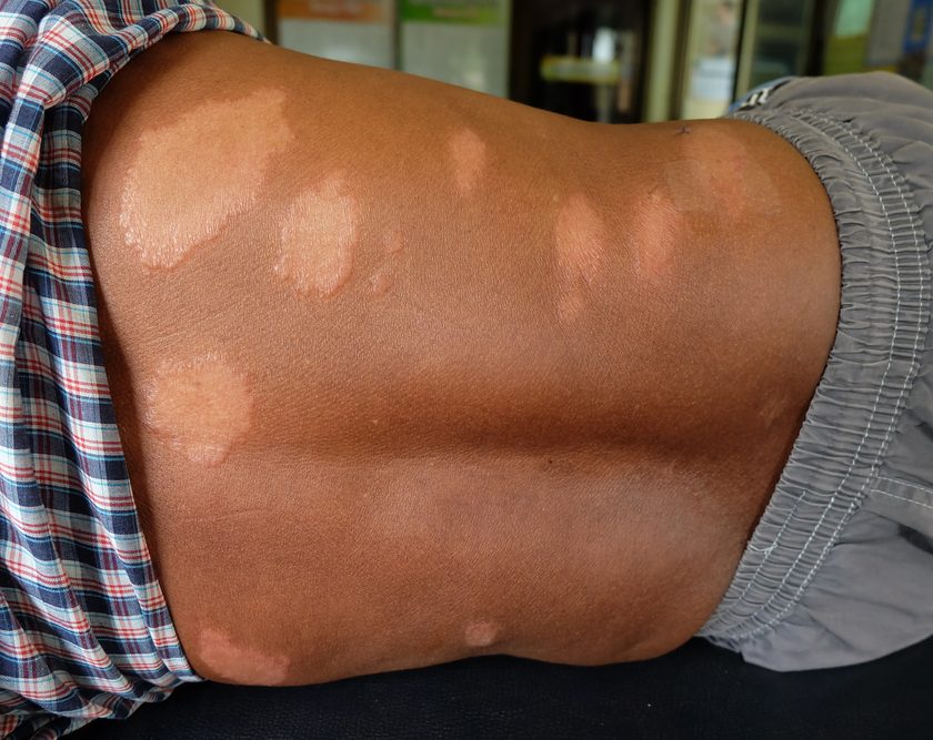
Infection-related peristomal complications include candidiasis and folliculitis. Peristomal candidiasis is an overgrowth of Candida organisms, with Candida albicans being the most common. Exposure to urine or fecal effluent provides a moist environment, which promotes the overgrowth of Candida organisms. The condition starts as pustules, which are abraded during pouch changes. Patients may complain of burning and itching. Treatment is aimed at keeping the peristomal skin dry and applying antifungal powder.
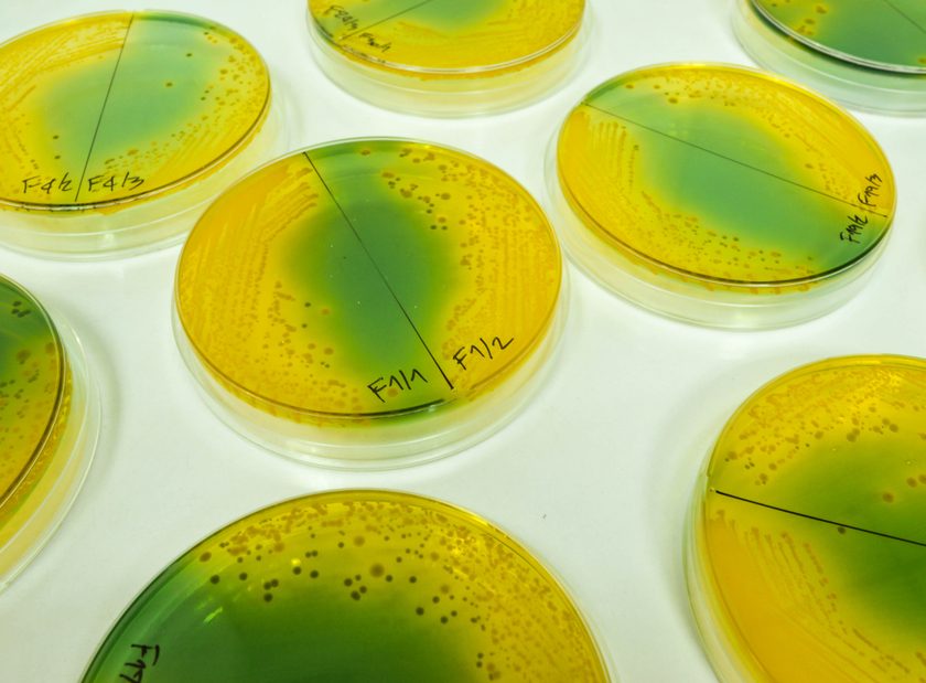
Folliculitis in the peristomal area is an inflammation of the hair follicles commonly due to shaving of the peristomal skin; it’s usually caused by Staphylococcus aureus. Prevention is key and involves clipping rather than shaving the skin, using antibacterial soap to cleanse the peristomal skin, gently removing the pouch, and using adhesive pouch-removal products to decrease the pulling of peristomal skin hairs when the pouch is removed.
M: Mechanical-related complications
Mechanical peristomal injuries can be related to pressure, friction, and epidermal stripping caused by the pouching system being too tight and rubbing against the peristomal skin. Other possible causes include traumatic removal of the pouch and too-aggressive cleansing of the peristomal skin during pouch changes. The peristomal skin may be erythematous or denuded or, in the case of pressure-related injuries, there may be a circumscribed ulcer.
Preventive measures include careful removal of the pouch, with gentle cleansing of the peristomal skin, or the use of a more flexible pouch if the pouching system rubs against the peristomal skin.
Once the injury has occurred, skin barrier powders may be applied over the denuded skin with a skin sealant. It’s important to reevaluate the pouching system to prevent mechanical injury from recurring.
E: Exposure of the peristomal skin to effluent
Exposure to effluent on the peristomal skin such as from an ileostomy can cause the skin to become erythematous in less than an hour, with skin breakdown in several hours. Urine can also cause problems because of the irritating effects of alkaline urine containing ammonium phosphates. Pseudoverrucous wartlike lesions may appear around urostomies that are chronically exposed to urine effluent, leading to thickening of the epidermis.
Use of chemical preparations (such as cleansers, liquid skin barriers, soaps, and adhesives) can also break down the peristomal skin. This type of skin breakdown is referred to as an irritant contact dermatitis; for example, if the soap used to clean the peristomal skin hadn’t been completely removed before the ostomy pouch was applied, the peristomal skin at the next pouch change may be erythematous due to the soap residue irritating the peristomal skin.
Some ostomates may develop an allergic contact dermatitis from hypersensitivity to certain chemicals in the ostomy products. Patch testing to determine which product is causing the allergen, then discontinuing the product, usually resolves the allergic dermatitis.
Treatment of exposure problems is aimed at finding the cause of the problem and establishing a secure pouching system that protects the peristomal skin from contact with the effluent or chemical preparation.
Be proactive
Unfortunately, many ostomates will experience peristomal skin complications. To proactively treat the signs of peristomal skin complications, clinicians and patients must be able to recognize them. Accurately describing the peristomal skin complication is important to determining which treatment works best for the ostomate and benchmarking treatment interventions that can be applied globally. Mnemonics such as DIME will help ensure that complications are caught early and patients receive the treatment they need.
All images courtesy of the Wound, Ostomy and Continence Nurses SocietyTM Image Library. Reprinted with permission.
Selected reference
Gray M, Colwell JC, Doughty D, et al. Peristomal moisture-associated damage in adults with fecal ostomies: a comprehensive review and consensus. J Wound Ostomy Continence Nurs. 40(4):389-399.
Catherine R. Ratliff is a clinical associate professor of nursing and program director of the Wound, Ostomy, and Continence Graduate Program at the University of Virginia School of Nursing in Charlottesville.
DISCLAIMER: All clinical recommendations are intended to assist with determining the appropriate wound therapy for the patient. Responsibility for final decisions and actions related to care of specific patients shall remain the obligation of the institution, its staff, and the patients’ attending physicians. Nothing in this information shall be deemed to constitute the providing of medical care or the diagnosis of any medical condition. Individuals should contact their healthcare providers for medical-related information.