About 1 million people in the United States have either temporary or permanent stomas. A stoma is created surgically to divert fecal material or urine in patients with GI or urinary tract diseases or disorders.
A stoma has no sensory nerve endings and is insensitive to pain. Yet several complications can affect it, making accurate assessment crucial. These complications may occur during the immediate postoperative period, within 30 days after surgery, or later. Lifelong assessment by a healthcare provider with knowledge of ostomy surgeries and complications is important.
Critical limb ischemia may not increase mortality risk in patients with diabetes Diabetic patients with critical limb ischemia (CLI) who are assessed quickly and treated aggressively do not have an increased risk of long-term cardiac mortality, according to a study in Diabetes Care.
By Nancy Chatham, RN, MSN, ANP-BC, CCNS, CWOCN, CWS, and Lori Thomas, MS, OTR/L, CLT-LANA An estimated 7 million people in the United States have venous disease, which can cause leg edema and ulcers. Approximately 2 to 3 million Americans suffer from secondary lymphedema. Marked by abnormal accumulation of protein-rich fluid in the interstitium, secondary lymphedema eventually can cause fibrosis…
By T. Michael Britton, RN, NHA, WCC, DWC As wound care professionals, we’ve all experienced a time when we felt that our patient didn’t have the appropriate wound treatment orders. However, the physician, nurse practitioner, or other prescriber wouldn’t follow your recommendation. This situation is not only frustrating but can delay the healing process. This article explores why a prescriber…
by Donna Sardina, RN, MHA, WCC, CWCMS, DWC, OMS What an honor it is to be the wound care “superhero”—the guru, the healer, the go-to person. Unfortunately, this honor may be accompanied by wound care overload—too much to do in too little time. Once someone is crowned the superhero specialist, others may try to transfer every aspect of wound and…
By Rosalyn S. Jordan, RN, BSN, MSc, CWOCN, WCC, OMS; and Judith LaDonna Burns, LPN, WCC, DFC About 1 million people in the United States have either temporary or permanent stomas. A stoma is created surgically to divert fecal material or urine in patients with GI or urinary tract diseases or disorders. A stoma has no sensory nerve endings and…
By Robyn Bjork, MPT, CWS, WCC, CLT-LANA The ability to understand or “read” lower-extremity redness in your patient is essential to determining its cause and providing effective treatment. Redness can occur in multiple conditions—hemosiderin staining, lipodermatosclerosis, venous dermatitis, chronic inflammation, cellulitis, and dependent rubor. This article provides clues to help you differentiate these conditions and identify the specific cause of…
By Donna Sardina, RN, MHA, WCC, CWCMS, DWC, OMS Knowledge is exploding online, making it essential that you’re comfortable using the Internet. You can also go online to save time and find a job, among other tasks. (See Online value.) However, you also need to keep in mind that anyone can put information on the Internet. As the caption of…
By Nancy Chatham, RN, MSN, ANP-BC, CCNS, CWOCN, CWS, and Lori Thomas, MS, OTR/L, CLT-LANA
An estimated 7 million people in the United States have venous disease, which can cause leg edema and ulcers. Approximately 2 to 3 million Americans suffer from secondary lymphedema. Marked by abnormal accumulation of protein-rich fluid in the interstitium, secondary lymphedema eventually can cause fibrosis and other tissue and skin changes. (more…)
At some point, most of us have encountered a bully—most commonly when we were kids. You might think that as we get older, bullying wouldn’t be a problem we have to deal with. Unfortunately, that’s not the case. In the healthcare field, bullying can be even worse than it was when we were children.
Bullying in health care takes many different forms, including fighting among different types of clinicians, managers bullying subordinates, peer-to-peer bullying and, most commonly, specialists bullying other specialists. Years ago when I realized my dream of becoming a wound care specialist, I thought other specialists would be relieved I was on board to help with the overwhelming task of spreading wound care knowledge and healing wounds. But I found out quickly that I was pretty much alone with those thoughts, and my first encounter with wound care bullies occurred.
I began to ask myself: What did I do wrong? Why are they slamming me? What did I do to them? They don’t even know me; they’ve never even talked to me. This may sound familiar to many of you, whether you’re a wound care specialist, an ostomy specialist, or a diabetes or lymphedema specialist.
Workplace bullying is defined as repeated, unreasonable actions by individuals (or a group) directed toward an employee (or group of employees) that are intended to intimidate, degrade, humiliate, or undermine. Bullying occurs for many reasons; these reasons almost always include insecurity, competition, and the desire to feel more powerful and be in control.
So how do we deal with the bullies?
• Follow the Golden Rule: Treat others as you’d like others to treat you. Don’t stoop to the bully’s level.
• Stay calm and rational. Don’t get emotional. Bullies take pleasure in manipulating people emotionally.
• Don’t lose your confidence or blame yourself. Recognize that this isn’t about you; it’s about the bully. Be proud and confident in your certification credential.
• Focus on your purpose—to provide safe, competent, high-quality care to every patient.
• Document the bullying incident. Start a diary detailing the nature of the bullying, including dates, times, places, what was said or done, and who was present. Start a file with copies of anything in print that shows harassment and bullying; hold onto copies of documents that contradict the bully’s accusations against you.
• If the bullying behavior compromises patient safety and care, report the bully.
Stopping all bullying in health care may seem like an insurmountable goal, but I believe that together we can try to stop the bullying cycle in our specialty. By setting the example and supporting each other, we can turn the focus back to healing and caring for our patients as a team, not as one practitioner against the world.
Actions speak louder than words. As Ralph Waldo Emerson said, “What you do speaks so loudly that I cannot hear what you say.”
Donna Sardina, RN, MHA, WCC, CWCMS, DWC, OMS
Editor-in-Chief Wound Care Advisor
Cofounder, Wound Care Education Institute
Plainfield, Illinois
By Rosalyn Jordan, RN, BSN, MSc, CWOCN, WCC, and Marci Christian, BBE
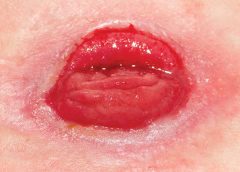
Any patient with a fecal or urinary ostomy may experience complications on the skin surface around the stoma. These complications may occur lifelong, although they’re more common during the first 5 years after the initial ostomy surgery. Causative factors include infection, trauma, certain diseases, and chemical irritation; most of these problems stem from the pouching system or pouch leakage.
Peristomal skin complications can cause a wide range of signs and symptoms, from skin discoloration to polyp-like growths, from erythema to full-thickness wounds. They can lead to discomfort, pain, poor self-image, social isolation, and impaired quality of life, not to mention additional care costs.
Incidence and types of these complications are hard to compare or contrast across multiple patients. Until recently, no standardized assessment or documentation tools were available to characterize or define complications. For this reason, reported rates ranged widely, from 10% to 70%. And because no designated common language or categories related to peristomal skin complications existed, documentation was inconsistent.
In the late 2000s, a group of nurses experienced in caring for ostomy patients worked with the World Council of Enterostomal Therapists to develop a resource called the Ostomy Skin Tool, which clinicians can use to categorize and describe peristomal skin complications in a consistent, objective manner. The tool also provides a common language for documentation.
The Ostomy Skin Tool has three major assessment domains—discoloration (D), erosion/ulceration (E), and tissue overgrowth (T), known collectively as DET. The DET combined rating ranges from normal, rated 0, to the worst condition possible, rated 15. Mild DET complications are documented as less than 4, moderate as less than 7, and severe as 8 or higher. (See Using the Ostomy Skin Tool by clicking the PDF icon above.)
The tool describes four categories of peristomal complications:
• chemical irritation
• mechanical trauma
• disease-related complications
• infection-related complications.
Chemical irritation
Chemical irritation can stem from irritants (as in contact dermatitis) or allergic reactions (allergic dermatitis). The most likely cause of chemical dermatitis is effluent leakage (feces or urine) from the colostomy, ileostomy, or urostomy, in which effluent comes in contact with peristomal skin. Other potential causes include contact with soap, certain adhesives, and adhesive removers.
The major treatment of chemical irritation is identification and removal of the offending agent, followed by patient and caregiver education on the new pouching procedure the patient must use. Follow-up assessment also is recommended. In a 2010 study that followed 89 patients for 1 year after ostomy surgery, about 50% of subjects experienced peristomal skin complications, most of them from pouch leakage. Another investigator estimated that 85% of ostomy patients experience pouch leakage at some time during their lives. Pouch leakage usually occurs when stool is extremely liquid (for instance, ileostomy effluent). Other causes of pouch leakage include wearing a pouch more than half full of effluent and abdominal contours that aren’t level. Besides changes in the pouching system, treatment may entail adding products to the pouching system or removing certain agents.
Some patients experience allergic dermatitis in reaction to products used in the pouching system (such as skin barriers, belts, pouch closures, or adhesives). However, allergic dermatitis is rare. One 2010 study suggested allergic reactions to these products occur in only about 0.6% of patients with peristomal skin irritation. Most major ostomy product manufacturers provide a patch test on request to help identify allergic conditions. Once the offending product is discontinued, allergic dermatitis should resolve rapidly.
Mechanical trauma
Mechanical trauma usually results from either the pouching system itself or its removal. It also may result from harsh or multiple skin-barrier removals, pressure from convex rings or pouches, and abrasive cleansing techniques. Some researchers believe the stronger the adhesive barrier and the more often a pouch is changed, the greater the risk of epidermal damage.
Mechanical trauma may present as a partial-thickness ulcer caused by pressure, shear, friction, tearing, or skin stripping. Patients with fragile skin are susceptible to mechanical trauma, so less aggressive pouching systems may be preferred for them. Of course, if the pouching system is changed, the patient or caregiver needs to learn about the new system.
Disease-related complications
Disease-related peristomal complications may be linked to preexisting skin conditions, such as psoriasis, eczema (atopic dermatitis), or seborrheic dermatitis. Hyperplasia also may occur. This overgrowth of cells, which may appear as gray or reddish brown pseudoverrucous lesions, usually is linked to urinary ostomies, although it can occur with fecal ostomies as well. Vinegar soaks are the recommended treatment, in addition to a change in the pouching system and corresponding patient education.
Occasionally, other disease-related complications occur, including primary adenocarcinoma of the peristomal skin and peristomal pyoderma gangrenosum, a painful and problematic condition that presents as peristomal ulcers. Ulcer borders are well-defined with a bluish purple coloration at the edges. Infection must be ruled out, as this condition usually is linked to an autoimmune condition. Treatment includes pain management and, in most cases, a topical corticosteroid. Crohn’s disease also may manifest as a peristomal skin ulcer.
Infection-related complications
Infection-related complications may be bacterial or fungal. Two common peristomal skin infections are folliculitis and Candida fungal infections. An infection of the hair follicle that causes pustules, folliculitis usually stems from traumatic hair pulling in the peristomal area during pouch removal. It may warrant a prescribed antibiotic, along with patient teaching regarding proper hair removal using an electric razor.
Candida infections may arise because peristomal skin provides a warm, dark, moist environment that promotes fungal growth. These infections appear as erythema with pustules or papules and satellite lesions. Treatment usually involves antifungal powder and use of the crusting technique to secure the pouching system. (See Using the crusting technique by clicking the PDF icon above.)
Management
Many complications are well advanced by the time patients seek assistance, perhaps because they don’t understand the significance of their symptoms and think they can manage the problem themselves. In some cases, they don’t know where to turn for assistance. Commonly, the complication progresses to the point where the patient goes to the emergency department or (particularly during the immediate postoperative period) needs to be readmitted for treatment. The best way to manage peristomal skin complications is to prevent them in the first place. (See Preventing peristomal skin complications by clicking the PDF icon above.)
Patient education
Over the past 20 years, hospital stays for ostomy surgery patients have decreased from about 2 weeks to less than 5 days. Reduced stays decrease the time available for caregivers to teach patients and family members how to empty and change the pouch. They need alternative education covering (among other topics) how to recognize peristomal skin complications and when to seek help. Not only do these complications require vigilant self-observation, but many patients don’t understand their implications or how rapidly they can worsen. In some cases, the first symptoms are itching and redness under the skin barrier. Fortunately, some patients may know or remember that itching, burning, stinging, reddened, or weeping peristomal skin requires professional attention. They can avoid serious complications by seeking assistance early, such as right after noticing pouch leakage.
Early treatment can reduce the cost of treatment. In a 2012 study, researchers estimated care costs related to peristomal skin complications for a 7-week treatment period, using the Ostomy Skin Tool as a reference. Severe complications (those with a DET score above 8) cost six times more to treat than mild cases (those with a DET score below 4) and 4.5 times more than moderate cases.
Along with early intervention by a trained ostomy care specialist, self-assessment by ostomy patients promotes a better quality of life, reduces pain, and may decrease care costs. Clinicians’ use of the Ostomy Skin Tool to assess and document peristomal skin complications promotes more reliable, objective, comparable assessment data for reporting.
Selected references
Al-Niaimi F, Lyon CC. Primary adenocarcinoma in peristomal skin: a case study. Ostomy Wound Manage. 2010;56(1):45-7.
Burch J. Management of stoma complications. Nurs Times. 2011;107(45):17-8, 20.
Jemec GB, Martins L, Claessens I, et al. Assessing peristomal skin changes in ostomy patients: validation of the Ostomy Skin Tool. Br J Dermatol. 2011; 164;330-5.
Jones T, Springfield T, Brudwick M, Ladd A. Fecal ostomies: practical management for the home health clinician. Home Healthc Nurse. 2011;29(5):306-17.
Martins L, Samai O, Fernandez A, et al. Maintaining healthy skin around an ostomy: peristomal skin disorders and self-assessment. Gastrointest Nurs. 2011;
9(2):9-13.
Martins L, Tavernelli K, Serrano JLC. Introducing a peristomal skin assessment tool: The Ostomy Skin Tool. World Council Enterostomal Therapists J. 2008;28(2):3-13.
Meisner S, Lehur P, Moran B, et al. Peristomal skin complications are common, expensive, and difficult to manage: a population based cost modeling study. PLoS One. 2012;7(5):e37813.
Omura Y, Yamabe M, Anazawa S. Peristomal skin disorders in patients with intestinal and urinary ostomies: influence of adhesive forces of various hydrocolloid wafer skin barriers. J Wound Ostomy Continence Nurs. 2010;37(3):289-98.
Ratliff CR. Early peristomal skin complications reported by WOC nurses. J Wound Ostomy Continence Nurs. 2010;37(5):505-10.
Shabbir J, Britton DC. Stomal complications: a literature overview. Colorectal Dis. 2010;12(10):958- 64.
Wound, Ostomy, Continence Clinical Practice Ostomy Subcommittee. Peristomal skin complications: Best practice for clinicians. Mt. Laurel, NJ; 2007.
The authors work for RecoverCare, LLC, in Louisville, Kentucky. Rosalyn Jordan is director of clinical education and Marci Christian is a clinical associate product specialist.
Assessing moisture and pressure risk in elderly patients continues to be a focus for clinicians in all settings, particularly long-term care. Ongoing research challenges our ideas about and practices for cleansing and protecting damaged skin. Until recently, most wound care clinicians have cleansed long-term care patients’ skin with mild soap and water. But several studies have shown pH-balanced cleansers are more efficient than soap and water for cleansing the skin of incontinent patients.
Various terms are used to describe skin breakdown related to moisture—incontinence-associated dermatitis, perineal dermatitis, diaper rash, intertriginal dermatitis, intertrigo, moisture-related skin damage, moisture-associated skin damage, and even periwound dermatitis. This article uses moisture-associated skin damage (MASD) because it encompasses many causes of skin breakdown related to moisture. Regardless of what we call the condition, we must do everything possible to prevent this painful and costly problem.
Skin assessment
Start with an overall assessment of the patient’s skin. Consider the texture and note dryness, flaking, redness, lesions, macerated areas, excoriation, denudement, and other color changes. (See Identifying pressure and moisture characteristics by clicking the PDF icon above.)
Assessing MASD risk
A patient’s risk of MASD can be assessed in several ways. Two of the most widely used pressure-ulcer risk scales, the Norton and Braden scales, address moisture risk. The Norton and Braden subscales should drive your plan for preventing skin breakdown related to moisture or pressure. The cause of breakdown (moisture, pressure, or shear/friction) must be identified, because treatment varies with the cause.
Both the Norton and Braden scales capture activity, mobility, and moisture scores. The Braden scale addresses sensory perception, whereas the Norton scale identifies mental condition. (See Subscales identifying pressure, shear, and moisture risk by clicking the PDF icon above.) Also, be aware that two scales have been published for perineal risk, but neither has been used widely.
You must differentiate pressure- and moisture-related conditions to determine correct treatment. Patients who are repositioned by caregivers are at risk for friction or shear. Also, know that agencies report pressure-ulcer prevalence. Care providers no longer classify mucous-membrane pressure areas in skin prevalence surveys; mucous membranes aren’t skin and don’t have the same tissue layers. Furthermore, don’t report skin denudement from moisture (unless pressure is present) in prevalence surveys.
When moisture causes skin breakdown
Skin has two major layers—epidermis and dermis. The epidermis itself has five layers: The outermost is the stratum corneum; it contains flattened, keratin protein–containing cells, which aid water absorption. These cells contain water-soluble compounds called natural moisturizing factor (NMF), which are surrounded by a lipid layer to keep NMF within the cell. When skin is exposed to moisture, its temperature decreases, the barrier function weakens, and skin is more susceptible to pressure and friction/shear injury. Also, when urea in urine breaks down into ammonia, an alkaline pH results, which may reactivate proteolytic and lipolytic enzymes in the stool. (See Picturing moisture and pressure effects by clicking the PDF icon above.)
Caring for moisture-related skin breakdown
The standard of care for moisture-related skin breakdown includes four major components: cleanse, moisturize, protect, and contain. Specific products used for each component vary with the facility’s product formulary.
Cleanse
Gently wash the area using a no-rinse cleanser with a pH below 7.0. Don’t rub the skin. Pat dry.
Moisturize
Use creams containing emollients or humectants. Humectants attract water to skin cells and help hold water in the cells; don’t use these products if the skin is overhydrated. Emollients slow water loss from skin and replace intracellular lipids.
Protect
Options for skin protectants include:
• liquid film-forming acrylate sprays or wipes
• ointments with a petroleum, zinc oxide, or dimethicone base
• skin pastes. Don’t remove these products totally at each cleansing, but do remove stool, urine, or drainage from the surface and apply additional paste afterward. Every other day, remove the paste down to the bare skin using a no-rinse cleanser or mineral oil.
Be sure to separate skinfolds and use products that wick moisture rather than trap it. These may include:
• commercial moisture-wicking products
• a light dusting with powder containing refined cornstarch or zinc oxide—not cornstarch from the kitchen or powder with talc as the only active ingredient
• abdominal pads.
Contain
To keep moisture away from skin, use absorbent underpads with wicking properties, condom catheters (for males), fecal incontinence collectors, fecal tubes (which require a healthcare provider order), or adult briefs with wicking or gel properties. Call a certified ostomy or wound care nurse for tips on applying and increasing wear time for fecal incontinence collectors.
If 4″ × 4″ gauze pads or ABD pads are saturated more frequently than every 2 hours, consider applying an ostomy or specially designed wound pouch to the area. Collecting drainage allows measurement and protects skin from the constant wetness of a saturated pad.
Don’t neglect the basics, for example, know that wet skin is more susceptible to breakdown. Turn the patient and change his or her position on schedule. Change linens and underpads when damp, and consider using a low-air-loss mattress or bed or mattress with microclimate technology.
Also, be aware that fungal rashes should be treated with appropriate medications. If the patient’s skin isn’t too moist, consider creams that absorb into the skin; a skin-protecting agent can be used as a barrier over the cream. Besides reviewing and using the standards of care, you may refer to the Incontinence-Associated Dermatitis Intervention Tool, which has categories related to skin damage. See the “Incontinence-Associated Dermatitis Intervention Tool” (IADIT).
Bottom line on skin breakdown
To help prevent skin breakdown related to moisture, assess patients’ skin appropriately, determine treatment using evidence-based guidelines, and implement an appropriate plan of care.
Selected references
Black JM, Gray M, Bliss DZ, et al. MASD part 2: incontinence-associated dermatitis and intertriginous dermatitis: a consensus. J Wound Ostomy Continence Nurs. 2011;38(4):359-70.
Borchert K, Bliss DZ, Savik K, Radosevich DM. The incontinence-associated dermatitis and its severity instrument: development and validation. J Wound Ostomy Continence Nurs. 2010;37(5):527-35.
Doughty D. Differential assessment of trunk wounds: pressure ulceration versus incontinence-associated dermatitis versus intertriginous dermatitis. Ostomy Wound Manage. 2012;58(4):20-2.
Doughty D, Junkin J, Kurz P, et al. Incontinence-associated dermatitis: consensus statements, evidence-based guidelines for prevention and treatment, and current challenges. J Wound Ostomy Continence Nurs. 2012;39(3):303-15.
Gray M, Beeckman D, Bliss DZ, et al. Incontinence-associated dermatitis: a comprehensive review and update. J Wound Ostomy Continence Nurs. 2012;
39(1):61-74.
Gray M, Black JM, Baharestani MM, et al. Moisture-associated skin damage: overview and pathophysiology. J Wound Ostomy Continence Nurs. 2011;38(3):233-41.
National Pressure Ulcer Advisory Panel and European Pressure Ulcer Advisory Panel. Prevention and treatment of pressure ulcers: clinical practice guideline.Washington, DC: National Pressure Ulcer Advisory Panel; 2009.
Wound, Ostomy and Continence Nurses Society. Guideline for Prevention and Management of Pressure Ulcers. Mt. Laurel, NJ: Wound, Ostomy and Continence Nurses Society; 2010.
Wound, Ostomy and Continence Nurses Society. Incontinence-Associated Dermatitis: Best Practice for Clinicians. Mt. Laurel, NJ: Wound, Ostomy and Continence Nurses Society; 2011.
Zulkowski K. Diagnosing and treating moisture-associated skin damage. Adv Skin Wound Care. 2012;25(5):231-6.
Patricia A. Slachta is an instructor at the Technical College of the Lowcountry in Beaufort, South Carolina.
Clinical practice guidelines help ensure we are applying the latest knowledge and expertise when we’re caring for patients. Here are a few recent guidelines that you may find useful.
Measurement of ankle-brachial index
An American Heart Association scientific statement, “Measurement and interpretation of the ankle-brachial index (ABI),” published in Circulation, outlines the use of ABI, terminology, how to calculate the value, training, standards, and suggestions for future research.
Recommendations for obtaining an ABI measurement include:
• Use the Doppler method to determine the systolic blood pressure in each arm and each ankle.
• Use the appropriate cuff size, with a width of at least 40% of the limb circumference.
• Place the ankle cuff just above the malleoli with the straight wrapping method.
• Cover open lesions with the potential for contamination with an impermeable dressing.
• Avoid using a cuff over a distal bypass.
The article also recommends measurement and interpretation of ABI be part of the standard curriculum for nursing and medical students. For more information about ABI, read “Bedside ankle-brachial index testing: Time-saving tips” in this issue of Wound Care Advisor.
Childhood obesity continues to be a significant problem in the United States, requiring innovative approaches for prevention and management. Those who are obese run the risk of poorer wound healing.
“Approaches to the prevention and management of childhood obesity: The role of social networks and the use of social media and related electronic technologies: A scientific statement from the American Heart Association,” published in Circulation, evaluates the role of social networks and social media in relation to childhood obesity and presents five steps for using social networks: 1 Define the goal of the intervention. 2 Identify the social network. 3 Develop and pilot test the intervention. 4 Implement the intervention. 5 Spread the intervention.
The guidelines conclude that social media holds promise as a tool, but more research is needed.
Guidelines for managing patients with stable ischemic heart disease
Many patients with wound or ostomy needs have comorbid heart disease. Be sure you are aware of the most current information for managing these patients by accessing “2012 ACCF/AHA/ACP/AATS/PCNA/SCAI/STS guideline for the diagnosis and management of patients with stable ischemic heart disease,” published in Circulation.
The guidelines acknowledge the vital importance of shared decision making between the healthcare provider and the patient. The information is divided into four sections with supporting algorithms:
• approaches to diagnosis
• risk assessment
• treatment
• follow-up.
Access the top 10 things to know and the executive summary.
In each case, the synthesis includes information in the following categories:
• areas of agreement and difference
• comparison of recommendations
• strength of evidence and recommendation grading schemes
• methodology
• source(s) of funding
• benefits and harms
• abbreviations
• status.
Access these and other guideline syntheses from AHRQ.
Bonus resource: Ethical case study of a patient refusing skin ulcer treatment
Free, one-time registration is required to view the entire video and all other
content on the Medscape website.
Patients have a right to make their own decisions, but what happens when a decision is so painful for staff that it affects morale? Arthur Caplan, PhD, Division of Medical Ethics at the NYU Langone Medical Center in New York, discusses such a case: “Patients have the right to choose death from bedsores.”
In most cases, amputation (removal of an extremity, digit, or other body part) is a surgical intervention performed to remove tissue affected by a disease and, in some cases, to provide pain relief. Fecal and urinary diversion surgeries also are considered amputations. Amputations and fecal or urinary diversions (ostomies) require extensive rehabilitation and adaptation to a new way of life, with physiologic and psychological impacts. Although diversions and ostomies usually are less visible to others than other types of amputations, they call for similar patient education, rehabilitatio n, and lifelong counseling.
The primary goal of therapy for ostomates and amputees is to resume their presurgical lifestyle to the greatest extent possible and to adapt to their new circumstances. Preoperative assessment and training interventions have proven valuable. Having a clear understanding of the surgical intervention helps reduce postoperative anxiety and depression, which can pose roadblocks to patients’ adaptation or response to their new situation. Successful interventions should be done by healthcare professionals who are trained in caring for ostomates and amputees.
Ostomates and amputees experience similar psychosocial challenges, body-
image problems, and sexuality concerns. This article focuses on these three issues. For a summary of other issues these patients may experience, see Other problems amputees and ostomates may face by clicking the PDF icon above.
Psychosocial challenges
Ostomates and amputees may experience depression, anxiety, fear, and many other concerns related to the surgical procedure—concerns that center on whether they’ll be able to resume their presurgical lifestyle. Many worry about social isolation and loss of income. Some fear both the primary disease process and the lifestyle changes induced by surgery. Anxiety may impede their social interactions and lead to significant psychological problems. Appropriate and effective counseling and therapy must be planned and provided. (But be aware that untrained or inexperienced healthcare professionals may not be able to provide the guidance the patient needs to feel comfortable; some may be unable even to offer information about available support systems.)
These patients also may find themselves socially isolated, in part due to loss of employment or the socioeconomic consequences of a decreased income. Some experience fear and worry when anticipating lifestyle changes caused by loss in or change of function, adaptation to the prosthesis, and treatment costs.
Maintaining social contact after surgery is extremely important to recovery and adaptation to the amputation or ostomy. The United Ostomy Associations of America and the Amputee Coalition encourage patients to maintain social involvement. Both groups suggest patients discuss their feelings, thoughts, and fears with a trusted family member, friend, or partner. Both organizations sponsor and encourage support-group involvement. In some cases, emotional support from other amputees or ostomates with a similar experience may be appropriate; some patients may be more comfortable sharing thoughts and asking questions in a group of people with similar experiences. Resuming presurgical social events and activities can enhance patients’ adaptation to a new way of life.
Ostomates and amputees have to cope not only with changes in physical appearance but with how their body functions and how they feel and perceive their body. They’re keenly aware of their changed appearance and are concerned about others’ perceptions of them. They may feel anxious and depressed related to body image; the degree of anxiety and depression may relate directly to their presurgical body image and activities. Many become anxious and fearful as they adapt to the prosthesis. (See Stages of grief by clicking the PDF icon above.)
Compared to amputees, ostomates may have more concerns about body image with sexual partners, because the stoma is, in a sense, a hidden amputation. In most cases, the stoma and pouch can be obscured visually from others. The amputee, on the other hand, has fewer options for hiding the missing body part.
To help patients cope with body-image problems, care providers must offer education, therapy, and counseling to help the patient accept and successfully adapt to the body-image change. The first step in this process may simply be to have the patient look at the stoma or stump, progressing to participation in prosthesis care.
Sexuality concerns
Many ostomates and amputees have difficulty resuming sexual activity after surgery. Although the stoma usually remains hidden from others, it’s observable to the ostomate and sex partner. Most patients require an adjustment period before they feel comfortable with a sex partner. They may fear that:
• the partner will reject them or no longer find them attractive
• they will experience loss of function and sensation
• they will experience pain or injury of the stoma.
They also may feel embarrassed, causing them to avoid sex. However, counselors can help couples discuss these concerns and resume a satisfactory sexual relationship. Ostomates and amputees and their partners may need counseling to resume a satisfactory sexual relationship. If they continue to have adjustment difficulties, referral to a trained sex counselor or psychologist may be indicated. Several studies show that appropriate counseling can help prevent complications and allow amputees and ostomates to continue to express their affection physically. (See Talking to patients about sexual problems by clicking the PDF icon above.)
Resuming sexual activity may be easier if the ostomate or amputee had a sex partner before surgery. However, males who experience postsurgical erectile dysfunction are less likely than other males to resume sexual activity. Counseling encourages postsurgical patients to focus more on the pleasurable feelings they and their partners feel, rather than on sexual performance. Body-image problems and inadequate sexual adjustment go hand in hand. (See Helping ostomates resume sex by clicking the PDF icon above.)
Team approach to patient education and counseling
In many parts of the country, a designated healthcare team manages amputees’ care and rehabilitation. But until recently, nurses were the only professionals certified to participate in ostomates’ care and rehabilitation. In fact, ostomates may represent a significant underserved population. A 2012 study found many ostomy patients didn’t receive consistent training and counseling from ostomy certified nurses. Only 13% of respondents reported they had regular visits with an ostomy certified nurse; 32% said they’d never received care from an ostomy nurse. Just over half (56%) indicated they saw an ostomy nurse when they thought it was necessary. The study also reported that 57% hadn’t seen an ostomy certified nurse in more than 1 year.
A team with specialized training to address ostomates’ physical and psychosocial needs might be able to provide the specialized care these patients need. The primary medical caregiver or general practitioner would serve as team leader and make appropriate referrals. The team should include a surgeon, ostomy- and amputee-trained nurses, a prosthetist or other healthcare provider trained in selection and fitting of prosthetic equipment and devices that affect function, a physical therapist, an occupational therapist, a social worker, a vocational counselor, a psychologist, caregiver or family members, support groups, and (last but not least) the patient.
The team approach might reduce hospital stays and promote patients’ return to their home environment. It also might encourage independence and enhance the success of long-term adaptation.
Focus on the future
Healthcare providers should encourage ostomates and amputees to focus on the future, not the past. Feeling comfortable with the prosthesis—the amputee’s artificial limb or the ostomate’s pouching system—is essential to adapting to a “new normal” way of life. Maintaining social relationships is important to adaptation as well. Mastering basic skills and adapting to changes in body function help improve the patient’s quality of life. Follow-up visits, phone contact, and access to a team of well-trained healthcare providers for patient education, rehabilitation, and long-term management are crucial to these patients’ successful adaptation and quality of life.
Selected references
Bhuvaneswar CG, Epstein LA, Stern TA. Reactions to amputation: recognition and treatment. Prim Care Companion J Clin Psychiatry. 2007;9(4):303-8.
Davidson T, Laberge M. Amputation. Gale Encyclopedia of Surgery: A Guide for Patients and Caregivers. 2004. www.encyclopedia.com/doc/1G2-3406200023.html. Accessed December 20, 2012.
Erwin-Toth P, Thompson SJ, Davis JS. Factors impacting the quality of life of people with an ostomy in North America: results from the Dialogue Study. J Wound Ostomy Continence Nurs. 2012;39(4):417-22.
Maguire P, Parkes CM. Surgery and loss of body parts. BMJ. 1998;316(7137):1086-8.
Pittman J, Kozell K, Gray M. Should WOC nurses measure health-related quality of life in patients undergoing intestinal ostomy surgery? J Wound Ostomy Continence Nurs. 2009;36(3):254- 65.
Pittman J. Characteristics of the patient with an ostomy. J Wound Ostomy Continence Nurs. 2011;38(3):271-9.
Racy JC. Psychological adaptation to amputation. In Bowker JH, Michael JW, ed. Atlas of Limb Prosthetics: Surgical, Prosthetic, and Rehabilitation Principles. 2nd ed. Rosemont, IL: American Academy of Orthopedic Surgeons; 1998.
Tunn PU, Pomraenke D, Goerling U, Hohenberger P. Functional outcome after endoprosthetic limb-salvage therapy of primary bone tumours—a comparative analysis using the MSTS score, the TESS and the RNL index. Int Orthop. 2008;32(5):619-25.
Turnbull G. Intimacy After Ostomy Surgery Guide. United Ostomy Associations of America, Inc. Revised 2009. www.ostomy.org. Accessed December 20, 2012.
Turnbull G. Sexuality after ostomy surgery. Ostomy Wound Manage. 2006;52(3):14,16.
A hot flush of embarrassment creates a bead of sweat on my forehead. “I’ve got to get this measurement,” I plead to myself. One glance at the clock tells me this bedside ankle-brachial index (ABI) procedure has already taken more than 30 minutes. My stomach sinks as I realize I’ll have to abandon the test as inconclusive. (more…)
January is traditionally the time of year when everyone starts the New Year fresh with resolutions for change—better organization, healthy living, new beginnings. This year, I want to challenge you to include your patients in your resolution planning. Work with each patient and come up with ideas to help improve their quality of life by, for instance, healing a wound, wearing a splint, keeping blood glucose levels within normal range, elevating the feet.
For resolutions to be effective, a “buy in” must exist. Most people don’t like
being told what they must do and would rather hear what they can do. So be sure to include your patients in the planning process. Ask them their opinion. Ask “What could I do differently to help you with this?” Determine if their treatments could be interfering with their lifestyle. Set mutually compatible goals. Most of all, help promote patients’ search for their own solutions.
To make an informed choice of can do instead of must do, patients must have a clear understanding of the facts, implications, and consequences. The Institute of Medicine defines health literacy as the degree to which individuals have the capacity to understand, obtain, and process basic health information needed to make appropriate health decisions. It’s easy to assume our patients know all the facts, especially if they have had the wound, ostomy, or diagnosis for many years. However, according to the 2003 National Assessment of Adult Literacy, almost 45% of the U.S. population (about 93 million Americans) have, at best, only basic health literacy skills.
To improve their health literacy, review your patients’ understanding of their situation and ask them to describe their diagnosis and treatment plan in their own words. If they can’t describe these correctly, reteach the information. Consider the possibility that you might have to use a new educational approach or method.
A new year and a new start, with a shared understanding of problems and potential solutions between the patient and wound care team can get you started on the pathway to success. To quote Zig Ziglar, “A goal properly set is halfway reached.”
Donna Sardina, RN, MHA, WCC, CWCMS, DWC, OMS
Editor-in-Chief Wound Care Advisor
Cofounder, Wound Care Education Institute
Plainfield, Illinois
NPUAP releases new position statement on exposed cartilage as Stage IV ulcer
The National Pressure Ulcer Advisory Panel (NPUAP) has released a new position statement, “Pressure ulcers with exposed cartilage are Stage IV pressure ulcers,” which states that pressure ulcers with exposed cartilage should be classified as Stage IV.
NPUAP notes that although the presence of “visible or palpable cartilage at the base of a pressure ulcer” wasn’t included in Stage IV terminology, cartilage “serves the same anatomical function as bone,” so it fits into the current Stage IV definition, “Full thickness tissue loss with exposed bone, tendon or muscle. Slough or eschar may be present on some parts of the wound bed. Often including undermining and tunneling.”
Medicare expenditures for diabetic foot care varies significantly by region
Medicare spending on patients with diabetes who have foot ulcers and lower extremity amputations varies significantly by region, according to a study in Journal of Diabetes and Its Complications, but more spending doesn’t significantly reduce 1-year mortality.
“Geographic variation in Medicare spending and mortality for diabetic patients with foot ulcers and amputations” examined data from 682,887 patients with foot ulcers and 151,752 patients with lower extremity amputations.
Macrovascular complications in patients with foot ulcers were associated with higher spending, and these complications in patients with amputations were more common in regions with higher mortality rates.
Rates of hospital admission were associated with higher spending and increased mortality rates for patients with foot ulcers and amputations.
“Geographic variation in Medicare spending and mortality rates for diabetic patients with foot ulcers and amputations is associated with regional differences in the utilization of inpatient services and the prevalence of macrovascular complications,” the study concludes.
Patients who develop pressure ulcers in hospital more likely to die
Medicare patients who develop pressure ulcers in the hospital are more likely to die during the hospital stay, have longer lengths of stay, and to be readmitted within 30 days after discharge, according to a study of 51,842 patients in the Journal of the American Geriatrics Society.
“Hospital-acquired pressure ulcers: results from the National Medicare Patient Safety Monitoring System Study” found that 4.5% of patients developed at least one new pressure ulcer during their hospitalization. Length of stay averaged 4.8 days for patients who didn’t develop a pressure ulcer, compared to 11.2 days for those with a new pressure ulcer.
Patients with diabetic foot ulcers may have higher risk of death
Patients with diabetes who have foot ulcers have a higher risk of cardiovascular disease and mortality, according to a meta-analysis in Diabetologia.
“The association of ulceration of the foot with cardiovascular and all-cause mortality in patients with diabetes: a meta-analysis” notes that the more frequent occurrence of cardiovascular disease only partly explains the increased mortality rate. Other explanations may include the more advanced stage of diabetes associated with those who had foot ulcers.
A Drugs.com article about the study reported that “analysis of data from more than 17,000 diabetes patients in eight studies found that the more than 3,000 patients with a history of foot ulcers had an extra 58 deaths per 1,000 people each year than those without foot ulcers.”
The study authors emphasize the importance of screening patients with diabetes for foot ulcers so intervention can begin early, as well as lowering cardiovascular risk factors.
Access patient information on foot care from the American Diabetes Association.
Nurse’s innovation for ostomy patients could improve quality of life
An oncology nurse in Australia has developed StomaLife, an alternative to ostomy bags.
StomaLife is a ceramic appliance that eliminates the need for an ostomy bag. According to the StomaLife website, the appliance uses a magnetic implant technology that provides a “pushing force” from within the body outward in order to keep the site intact, while a second part is placed on the stoma site. A cotton gauze pad is used between the skin and the appliance to keep the site separated and to provide air circulation to the surrounding skin.
“The benefits of StomaLife to ostomy patients are continence all day, reduced skin irritation and infection, odour and sound control, leak prevention, waste material flow control and on-demand gas release,” says Saied Sabeti.
StomaLife still needs to be tested and is not yet being produced.
New laser-activated bio-adhesive polymer aims to replace sutures
The Journal of Visualized Experiments, a peer-reviewed video journal, has published “A chitosan based, laser activated thin film surgical adhesive, ‘SurgiLux’: preparation and demonstration.”
SurgiLux is a laser-activated, bio-adhesive polymer that is chitosan-based. Chitosan is a polymer derived from chitin, which is found in fungal cell walls or in exoskeletons of crustaceans and insects. This molecular component allows SurgiLux to form low-energy bonds between the polymer and the desired tissue when it absorbs light.
The technology may be able to replace traditional sutures in the clinical setting. SurgiLux polymer can achieve a uniform seal when activated by a laser and has antimicrobial properties, which help prevent a wound from becoming infected. It also maintains a barrier between the tissue and its surroundings.
SurgiLux has been tested both in vitro and in vivo on a variety of tissues, including nerve, intestine, dura mater, and cornea.
Palliative care raises patient satisfaction and reduces costs
Kaiser Permanente’s home-based palliative care program increased patient satisfaction and decreased emergency department visits, inpatient admissions, and costs, according to an innovation profile in the Agency for Healthcare Research and Quality’s Innovations Exchange.
“In-home palliative care allows more patients to die at home, leading to higher satisfaction and lower acute care utilization and costs” notes that the program uses an interdisciplinary team of providers to manage symptoms and pain, provide emotional and spiritual support, and educate patients and family members on an ongoing basis about changes in the patient’s condition.
Other components of the program include a 24-hour nurse call center, biweekly team meetings, and bereavement services to the family after the patient dies.
More research needed to determine efficacy of maggot debridement therapy
“The efficacy of maggot debridement therapy (MDT)—a review of comparative clinical trials” concludes that “poor quality of the data used for evaluating the efficacy of MDT highlights the need for more and better designed investigations.”
The authors of the article in International Wound Journal reviewed three randomized clinical trials and five nonrandomized clinical trials evaluating the efficacy of sterile Lucilia sericata applied on ulcers.
The studies found that MDT was “significantly more effective than hydrogel or a mixture of conventional therapy modalities, including hydrocolloid, hydrogel and saline moistened gauze,” but the designs of the study were “suboptimal.”
Use tool to select correct antimicrobial dressing
“Ensuring that the correct antimicrobial dressing is selected,” in Wounds International, emphasizes that dressing selection should be based on assessment of the microbial burden in the wound, the wound type, and the location and condition of the wound.
The article includes a checklist that may be helpful for deciding on the level of bacterial burden in a wound. The checklist is used to determine four levels of risk—colonized: at risk; localized infection; spreading infection; and systemic infection. Each level has a corresponding definition.
A table of antimicrobial dressings reviews the antimicrobial agent and dressing form, and the article ends with a case study.
Necrotizing fasciitis (NF) results from an infection that attacks the fascia and subcutaneous tissues. The primary bacterial etiology is group A streptococcus, a facultative anaerobic bacterium. However, other bacteria may contribute. Sometimes called the “flesh-eating” disease because of the potentially devastating effect on the afflicted patient, NF can be monomicrobial or polymicrobial.
The four typical settings for NF are:
surgical bowel or abdominal trauma surgery
pressure ulcer and perianal abscess
injection sites (especially in drug users)
Bartholin abscess or minor vulvovaginal infection.
Because of the rapid course and ravaging nature of acute NF, clinicians must maintain a high index of suspicion if the patient has suggestive signs and symptoms. In 1990, puppeteer Jim Henson (best known for creating the Muppets) died from NF. At that time, little was known about the progression of group A streptococcal infection.
The disease can quickly cause death, so starting immediate treatment is even more crucial than confirming the diagnosis. Once the disease is suspected, antibiotics must be given immediately and the patient must be prepared for surgery at once. NF spreads rapidly, capable of progressing from a small lesion to death in days to weeks. Thus, delayed diagnosis increases the risk of death. Lack of knowledge about the disease and inability to recognize it promptly are the main reasons many victims die. This article can improve your knowledge base.
Overview
NF was discovered in 1871 by Joseph Jones, a Confederate Army surgeon. At that time, it was called hemolytic streptococcal gangrene, nonclostridial gas gangrene, nonclostridial crepitant cellulitis, necrotizing or gangrenous erysipelas, necrotizing cellulitis, bacterial synergistic gangrene, or synergistic necrotizing cellulitis.
NF involves the fascia, muscle compartments, or both. It can affect not only the muscle fascia but the superficial fascia. NF and cellulitis differ in the amount of tissue involved and extent of tissue involvement.
The most common areas of infection are the abdominal wall, perineum, and extremities. When NF affects the perineum and scrotum, it’s called Fournier gangrene, after the French dermatologist and virologist Alfred Jean Fournier.
The most common causes are trauma, surgery, and insect bites. The disease can affect persons of any age. Such comorbidities as diabetes, chronic renal failure, immunosuppressive therapy, hypertension, obesity, and malnutrition increase susceptibility.
Pathophysiology
NF falls into four classifications based on wound microbiology. Type 1, the most common, involves polymicrobial bacteria. Type 2 results from trauma and is associated with comorbidities. Type 3, rare in this country, stems from gram-negative marine bacteria. Type 4 is a fungal infection occurring mostly in immunocompromised persons. (See Comparing types of necrotizing fasciitis by clicking the PDF icon above.)
Disease progression
The four types of NF progress in a similar way. Bacteria secrete pyrogenic exotoxin A, which stimulates cytokines. These cytokines damage the endothelial lining; fluid then leaks into the extravascular space.
M proteins in streptococci and β-hemolytic streptococci exacerbate the immune reaction by inhibiting phagocytosis of polymorphonuclear leukocytes and normal neutrophil chemotaxis. As the immune reaction increases, blood vessels dilate, allowing toxins to leak through vessel walls, which in turn decreases blood flow. As the cascade continues, hypoxic conditions cause facultative aerobic organisms to grow and become anaerobic. These bacteria exacerbate destruction of surrounding cells and lead to release of carbon dioxide, water, hydrogen, nitrogen, hydrogen sulfide, and methane. As the infection continues to progress, toxins spread throughout the bloodstream and the patient becomes septic.
Assessment
Obtain the patient’s medical history and description of the wound. Determine when the changes first appeared and whether the affected area seemed to get worse recently.
In all NF types, patients commonly present with a small, painful area (possibly with entry marks) but no other signs or symptoms. The wound may appear as a bulla, cellulitis, or dermatitis, representing an infection developing in underlying tissues. The skin may have a wooden-hard feel as the infection progresses to the subcutaneous space and causes necrosis. The wound becomes discolored and necrotic; drainage is rare until surgical debridement begins. The patient quickly develops fever, chills, nausea, and vomiting. As NF progresses, bullae become dark purple with darkened edges; the patient grows disoriented and lethargic, and organ failure and respiratory failure
ensue. Without treatment, the patient dies.
Diagnosis
Diagnostic tests usually include magnetic resonance imaging, complete blood count with differential, comprehensive metabolic panel, and cultures. (See Diagnostic findings in necrotizing fasciitis by clicking the PDF icon above.)
Treatment
Immediate surgical debridement and broad-spectrum antibiotics are needed to stop the immune response to infection. Clindamycin, gentamicin, penicillin, or metronidazole may be given alone or in combination until culture results are available. Supportive care includes total parenteral nutrition for nutritional support, I.V. fluids, and oxygen. Limb amputation should be done only as a last resort.
Surgical debridement involves penetrating deep into the fascia and removing all necrotic tissue. After the first debridement, release of “dishwater fluid” may occur.
Administering hyperbaric oxygen therapy (HBOT) after the first debridement increases tissue oxygenation, thus reducing tissue destruction by anaerobic bacteria. During HBOT (usually given as a 90-minute treatment), the patient breathes 100% oxygen in an environment of increasing atmospheric pressure.
HBOT should be given in conjunction with surgical debridement (usually after each debridement) and should continue until necrotic tissue ceases and cell destruction stops. HBOT also promotes collagen synthesis and neoangiogenesis (new blood vessel growth), which boosts blood supply and oxygen to tissues.
Adverse effects of HBOT include ear pain, oxygen toxicity, and seizures. Ear pain can be minimized by swallowing or yawning. If the patient continues to have ear pain, ear tubes may be inserted by an otolaryngologist. During HBOT, air breaks (intervals of breathing room air) are important in controlling oxygen toxicity (the main cause of seizures).
Throughout the HBOT treatment period, wound dressings must be simple. Well-moistened gauze dressings and an abdominal pad provide good support. Once necrotic destruction occurs, dressings depend on wound size and the need to fill cavities. The patient may require a diverting colostomy, depending on wound
location and the amount of uncontrolled diarrhea. Blood glucose levels must be monitored before and after HBOT, as this treatment affects blood glucose.
Supportive care and follow-up treatment
During initial treatment, patients need supportive care and monitoring. Once they’re out of danger, begin teaching them how to prevent NF recurrences. Advise them to control blood glucose levels, keeping the glycated hemoglobin (HbA1c) level to 7% or less. Caution patients to keep needles capped until use and not to reuse needles. Instruct them to clean the skin thoroughly before blood glucose testing or insulin injection, and to use alcohol pads to clean the area afterward.
Before discharge, help arrange the patient’s aftercare, including home health care for wound management and teaching, social services to promote adjustment to lifestyle changes and financial concerns, and physical therapy to help rebuild strength and promote the return to optimal physical health. One helpful patient resource is the National Necrotizing Fasciitis Foundation. The Centers for Disease Control and Prevention section on necrotizing fasciitis includes “Common sense and great wound care are the best ways to prevent a bacterial skin infection.”
The life-threatening nature of NF, scarring caused by the disease, and in some cases the need for limb amputation can alter the patient’s attitude and viewpoint, so be sure to take a holistic approach when dealing with the patient and family. Today, NF has a much better survival rate than 2 decades ago when Jim Henson died. In my practice, I’ve seen four NF cases. Thanks to early identification, good wound care, and HBOT, these patients suffered only minimal damage.
Selected references
Boyer A, Vargas F, Coste F, et al. Influence of surgical treatment timing on mortality from necrotizing soft tissue infections requiring intensive care management. Intensive Care Med. 2009;35(5):847-853. doi:10.1007/s00134-008-1373-4.
Cain S. Necrotizing fasciitis: recognition and care. Practice Nurs. 2010;21(6):297-302.
Centers for Disease Control and Prevention. Notes from the field: fatal fungal soft-tissue infections after a tornado—Joplin, Missouri, 2011. MMWR. 2011;60(29):992.
Chamber AC, Leaper DJ. Role of oxygen in wound healing: a review of evidence. J Wound Care. 2011; 20(4):160-164.
Christophoros K, Achilleas K, Vasilia D, et al. Postraumatic zygomycotic necrotizing abdominal wall fasciitis with intraabdominal invasion in a non immunosuppressed patient. Internet J Surg. 2007;11(1). doi:10.5580/17a8.
Ecker K-W, Baars A, Topfer J, Frank J. Necrotizing fasciitis of the perineum and the abdominal wall-surgical approach. Europ J Trauma Emerg Surg. 2008;
34(3):219-228. doi:10.1007/s00068-008-8072-2.
Hunter J, Quarterman C, Waseem M, Wills A. Diagnosis and management of necrotizing fasciitis. Br J Hosp Med. 2011;72(7):391-395.
Magel DC. The nurse’s role in managing necrotizing fasciitis. AORN J. 2008;88(6):977-982.
Phanzu MD, Bafende AE, Imposo BB, Meyers WM, Portaels F. Under treated necrotizing fasciitis masquerading as ulcerated edematous Mycobacterium ulcerans infection (Buruli ulcer). Am J Trop Med Hyg. 2012;82(3):478-481.
Ruth-Sahd LA, Gonzales M. Multiple dimensions of caring for a patient with acute necrotizing fasciitis. Dimens Crit Care Nurs. 2006;25(1):15-21.
Stevens DL, Bisno AL, Chambers HF, et al; Infectious Diseases Society of America. Practice guidelines for the diagnosis and management of skin and soft-tissue infections. Clin Infect Dis. 2005;41(10):1373-1406.
Su YC, Chen HW, Hong YC, Chen CT, et al. Laboratory risk indicator for necrotizing fasciitis score and the outcomes. ANZ J Surg. 2008;78(11):968-972.
Taviloglu K, Yanar H. Necrotizing fasciitis: strategies for diagnosis and management. World J Emerg Surg. 2007;2:19.
Lydia Meyers is a medical reviewer for National Government Services in Castleton, Indiana, and a clinical liaison at CTI Nutrition in Indianapolis. She has 11 years of wound care experience in nursing homes, wound clinics, and home health.
Complex wound failures are costly and time-consuming. They increase length of stay and contribute to morbidity and mortality in surgical patients. Negative-pressure wound therapy (NPWT)—a common adjunct to wound-care therapy—is used to accelerate wound healing in all fields of surgery. Using a vacuum device and wound-packing material, it applies subatmospheric pressure to complex wounds.
But NPWT alone doesn’t ensure adequate wound healing. Many physiologic factors—including infection, excessive moisture, nutrition, and medications—influence wound-healing success. Failure to account for these factors or improper application of NPWT can limit patient outcomes and cause debilitating complications.
For clinicians, applying and establishing an airtight seal on a complex wound is among the most dreaded, time-consuming, and challenging NPWT-related tasks. Simply applying NPWT material under layers of transparent drape may delay wound healing or exacerbate the wound. This article provides tips on safe application of NPWT to enhance the outcomes of patients with complex wounds.
Consider wound location
Wounds on the body’s anterior surfaces are less susceptible to the forces of pressure, friction, and shear than those on posterior and lateral surfaces. Posterior and lateral wounds commonly require posterior offloading or repositioning the patient in bed to reduce or eliminate direct pressure. This can be done with judicious and frequent patient turning using a specialty bed or support surface.
Bridge a posterior or lateral wound to an anterior surface by placing the drainage collection tubing to a nonpressure-bearing surface away from the wound. Bridging keeps the tubing from exerting pressure on intact skin and decreases the risk of a pressure ulcer. To create the bridge, cut foam into a single spiral of 0.5 to 1 cm, or if using gauze, fold gauze into 8 single layers.
Place the spiraled foam or gauze layers onto the drape, ensure the bridge is wider than the collection tubing disc, and secure it with an additional drape. Next, apply the NPWT collection tubing on the end of the bridge away from the wound. A wide bridge under the collection tubing disc will minimize the potential for periwound breakdown when negative pressure is initiated. You may modify this spiraling technique by varying the width of the foam to fill undermining and wounds of irregular configuration and depth.
Protect the periwound
An intact periwound may break down from exposure to moisture, injury from repetitive removal of a transparent drape, or NPWT material coming in contact with skin. Skin protection is critical in preventing additional breakdown stemming from contact with potentially damaging material.
Transparent drapes are designed to permit transmission of moisture vapor and oxygen. Avoid using multiple layers of transparent drapes to secure dressings over intact skin, as this can decrease the transmission of moisture vapor and oxygen, which in turn may increase the risk of fungal infection, maceration, and loss of an intact seal.
Periwound maceration also may indicate increased wound exudate, requiring an increase in negative pressure. Conversely, an ecchymotic periwound may indicate excessively high negative pressures. If either occurs, assess the need to adjust negative pressure and intervene accordingly. Reassess NPWT effectiveness with subsequent dressing changes.
Apply a protective liquid skin barrier to the periwound and adjacent healthy tissue to help protect the skin surface from body fluids. The skin barrier also helps prevent stripping of fragile skin by minimizing shear forces from repetitive or forceful removal of transparent drapes. Excessive moisture can be absorbed by using a light dusting of ostomy powder sealed with a skin barrier. A “window pane” of transparent drape or hydrocolloid dressing around the wound also can protect surface tissue from contactwith NPWT material and prevent maceration.
Avoid creating rolled wound edges
In the best-case scenario, epithelial tissue at the wound edge is attached to the wound bed and migrates across healthy granulation tissue, causing the wound to contract and finally close. With deep wound environments that lack moisture or healthy granulation tissue, the wound edges may roll downward and epibole may develop. Epibole is premature closure of the wound edges, which prevents epithelialization and wound closure when it comes in contact with a deeper wound bed. (See Picturing epibole by clicking the PDF icon above.)
Materials used in NPWT are primarily air-filled. Applying negative pressure causes air removal, leading to wound contraction by pulling on the wound edges—an action called macrostrain. Without sufficient NPWT material in the wound, macrostrain can cause the wound to contract downward and the wound edges to roll.
Ensure that enough NPWT material has been applied into the wound to enhance wound-edge approximation and avoid creating a potential defect as the wound heals. Before NPWT begins, material should be raised 1 to 2 cm above the intact skin. Additional material may be needed with subsequent changes if the NPWT material compresses below the periwound. The amount of NPWT material needed to remain above the periwound once NPWT starts varies with the amount of material compressed and the wound depth.
Reduce the infection risk
To some degree, all wounds are contaminated. Usually, the body’s immunologic response is able to clear bacterial organisms and wound healing isn’t delayed. But a patient who has an infection of a complex wound needs additional support.
Systemic antibiotics alone aren’t enough because they’re selective for specific organisms and don’t reach therapeutic levels in the wound bed. In contrast, topical antimicrobial adjuncts, such as controlled-release ionic silver, provide broad-spectrum antimicrobial coverage against fungi, viruses, yeasts, and gram-negative and gram-positive bacteria, including methicillin-resistant Staphylococcus aureus and vancomycin-resistant enterococci.
Consider using controlled-release ionic silver for a wound known to be infected or at risk for infection due to its location or potential urine or fecal contamination. To be bactericidal, ionic silver must be in concentrations of at least 20 parts per million; also, it must be kept moist and must come in direct contact with infected wound bed. At lower concentrations, organisms may develop resistance. Ionic silver has no known resistance or contraindications. Dressings using it come in several forms, including a hydrogel sheet, perforated sheet, cavity version, and semiliquid hydrogel. Be sure the form you choose doesn’t occlude the NPWT material and compromise therapy. (See NPWT for a patient with necrotizing fasciitis by clicking the PDF icon above.)
One of the most daunting aspects of NPWT is obtaining and maintaining a good seal—in other words, avoiding the dreaded leak. Preventive skin measures may contribute to a poor seal; skin-care products containing glycerin, surfactant, or dimethicone may prevent adequate adhesion of NPWT drapes. Body oil, sweat, and hair may need to be minimized or removed.
To avoid leaks, don’t overlook the obvious—loose connections, a loose drainage collection canister, exposed NPWT material, and skinfolds extending beyond the transparent drape. Tincture of benzoin (with or without a thin hydrocolloid dressing) increases tackiness to enhance the adhesive property of a transparent drape on the diaphoretic patient and on hard-to-drape areas, such as the perineum. But be sure to use tincture of benzoin with discretion, as it may remove fragile periwound tissue when the dressing is removed.
Ostomy paste products can serve as effective filler. These pliable products can be spread into position to obtain a secure seal under the transparent drape in hard-to-seal areas, such as the perineum. Pastes remain flexible and can be removed without residue. Temporarily increasing NPWT pressure to a higher setting may help locate a subtle leak or provide enough negative pressure to self-seal the leak. Once the leak resolves, remember to return the pressure to the ordered setting.
Knowledge optimizes healing
It’s important to be aware of potential complications of NPWT (See Take care with NPWT by clicking on the PDF icon above). However, when applied correctly, NPWT is an effective option for managing complex wounds. Recognizing and managing potential complications at the wound site, ensuring periwound protection, minimizing epibole formation, and preventing wound infection can result in a better-prepared wound bed and promote optimal healing.
Selected references
Baranoski S, Ayello EA. (2012). Wound Care Essentials: Practice Principles. 3rd ed. Springhouse, PA; Lippincott Williams & Wilkins.
Bovill E, Banwell PE, Teot L, et al. Topical negative pressure wound therapy: a review of its role and guidelines for its use in the management of acute wounds. Int Wound J. 2008;5:511-529.
Sussman C, Bates-Jensen B. Wound Care: A Collaborative Practice Manual for Health Professionals. 4th ed. Baltimore, MD; Lippincott Williams & Wilkins; 2011.
Ronald Rock is an Adult Health Clinical Nurse Specialist in the Digestive Disease Institute at the Cleveland Clinic in Cleveland, Ohio.