By Nancy Morgan, RN, BSN, MBA, WOC, WCC, DWC, OMS
Each month, Apple Bites brings you a tool you can apply in your daily practice.
Description
Xerosis, an abnormal dryness of the skin, is one of the most common skin conditions among patients with type 2 diabetes. While assessing for predictors of foot lesions in patients with diabetes, the authors of one study found that 82.1% of these patients had skin with dryness, cracks, or fissures. An unpublished survey of 105 consecutive patients with diabetes revealed that 75% had clinical manifestations of dry skin.
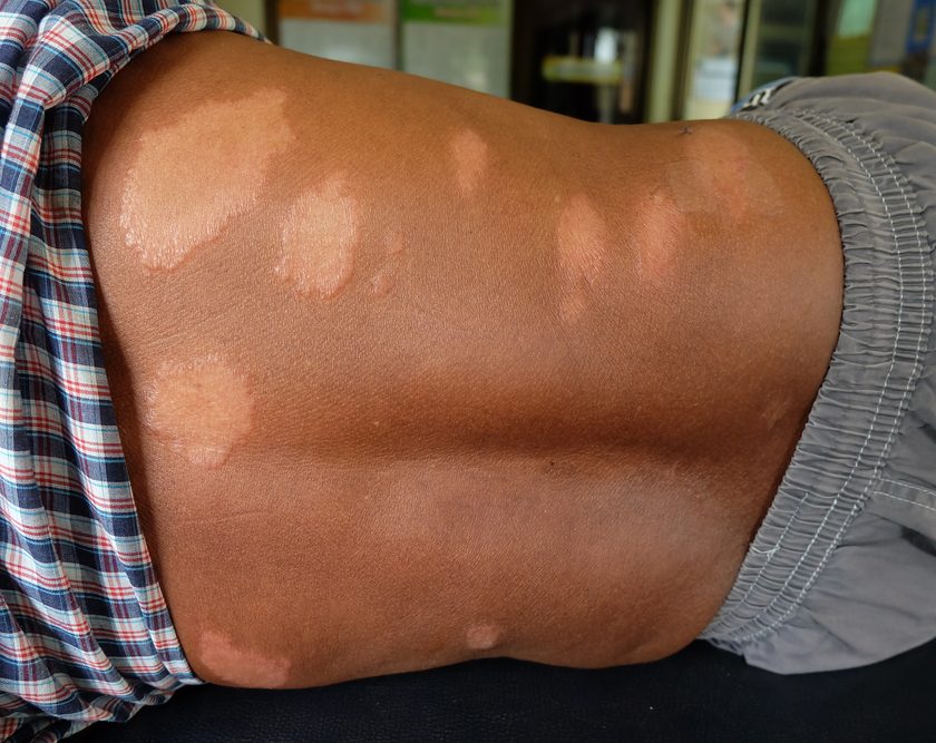
Characteristics
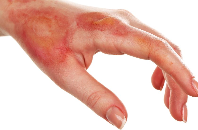
• Excessively dry, rough, uneven, and cracked skin
• Possible raised or uplifted skin edges (scaling), desquamation (flaking), chapping, and pruritus
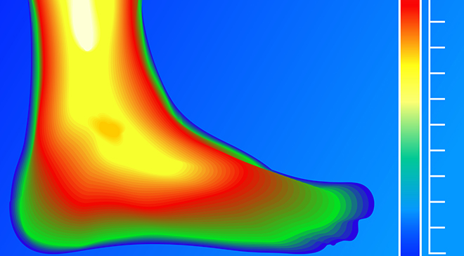
• Most common on the heels and feet
• Can lead to fissures (linear cracks in the skin) with hyperkeratotic tissue
Progression
The progression of xerosis follows a defined pattern:
• Initially, the skin becomes dry and rough, with pronounced skin lines.
• As the condition progresses, superficial scaling with fissuring and erythema develops. In severe cases, a crisscrossing pattern with superficial scaling is present.
• The skin becomes less elastic and
loses both its flexibility and its ability to withstand trauma, which may result in skin breakdown and subsequent infection.
Causes
• There is a loss of natural moisturizing factors and moisture from the stratum corneum and intercellular matrix of the skin.
• Sebaceous and sweat glands normally maintain skin lubrication and control the oil and moisture in the foot, but they become atrophied when autonomic neuropathy occurs.
• Corneocytes are aligned parallel to each other in normal skin; xerosis causes structural changes to these cells and disrupts the surface, resulting in a rough epidermal surface.
• The dryness is due to the redistribution of blood flow in the soles of the feet by persistent and inappropriate dilatation of arteriovenous shunts. This activity diverts blood away from the skin surface. When this occurs in combination with alterations in the elasticity of the skin (due to nonenzymatic glycosylation of structural proteins and glycoproteins), the skin splits, creating portals for bacteria to enter.
TEST your general knowledge about xerosis (questions 14 to 24).
Treatment
Apply an agent to maintain skin moisture, such as an emollient lotion or cream, to the feet daily. Use moisturizers that contain urea or lactic acid.
• Urea works by enhancing the water-binding capacity of the stratum corneum. Long-term treatment with urea has been demonstrated to decrease transepidermal water loss. Urea also is a potent skin humidifier and descaling agent, particularly in 10% concentration.
• Lactic acid (in the form of an alpha hydroxy acid) can accelerate softening of the skin, dissolving or peeling the outer layer of the skin to help maintain its capability to hold moisture. Lactic acid in concentrations of 2.5% to 12% is the most common alpha hydroxy acid used for moderate to severe xerosis.
• Examples of products with urea or lactic acid include Atrac-Tain Cream, Eucerin 10% Urea Lotion, Lac-Hydrin 12%, and AmLactin Foot Cream Therapy.
It’s important to avoid:
• products that contain alcohol because they evaporate and their drying action compounds the original problem.
• petroleum-based products, which seal the skin surface and prevent what little lubrication made from evaporating. These products don’t penetrate the surface of the skin and don’t replace skin moisture.
Patient education
Tell patients with xerosis to:
• minimize bathing to no more than once a day or even every other day
• use cool or lukewarm water
• pat, don’t rub, to dry the skin
• avoid harsh soaps
• avoid lotions with dyes or perfumes.
Also explain how to apply—and how often to apply—skin moisturizers.
Note: Clinicians should routinely inspect the feet of patients with diabetes.
Selected references
Flynn TC, Petros J, Clark JE, et al. Dry skin and moisturizers. Clin Dermatol. 2001;19(4):387-392.
Hill MJ. Fungal infections. Dermatol Nurs. 2008; 20:137-138.
Litzelman DK, Marriott DJ, Vinicor F. Independent physiological predictors of foot lesions in subjects with NIDDM. Diabetes Care. 1997;20(8):1273-1278.
Pham HT, Exelbert L, Segal-Owens AC, Veves A. A prospective, randomized, controlled double-blind study of a moisturizer for xerosis of the feet in patients with diabetes. Ostomy Wound Manage. 2002;48(5):30-36.
Rehm K. Towards better management of diabetic foot complications. Podiatry Manage. 2007;26(9):243-254.
Serrup J. A three-hour test for rapid comparison of effects of moisturizers and active constituents (urea). Measurement of hydration, scaling, and skin surface lipidization by non-invasive techniques. Acta Derm Venereol Suppl. (Stockh). 1992;177:29-33.
Smith RG. A guide to skin conditions of the diabetic foot. Podiatry Today. 2004;17(9):48-58.
Nancy Morgan, cofounder of the Wound Care Education Institute, combines her expertise as a Certified Wound Care Nurse with an extensive background in wound care education and program development as a nurse entrepreneur. Read her blog, “Wound Care Swagger.”
Information in Apple Bites is courtesy of the Wound Care Education Institute (WCEI), copyright 2013.