Flesh-eating bacteria infections are on the rise in the US

Vibrio vulnificus

By Bill Sullivan, Indiana University

Flesh-eating bacteria sounds like the premise of a bad horror movie, but it’s a growing – and potentially fatal – threat to people.

In September 2023, the Centers for Disease Control and Prevention issued a health advisory alerting doctors and public health officials of an increase in flesh-eating bacteria cases that can cause serious wound infections.

I’m a professor at the Indiana University School of Medicine, where my laboratory studies microbiology and infectious disease. Here’s why the CDC is so concerned about this deadly infection – and ways to avoid contracting it.

What does ‘flesh-eating’ mean?

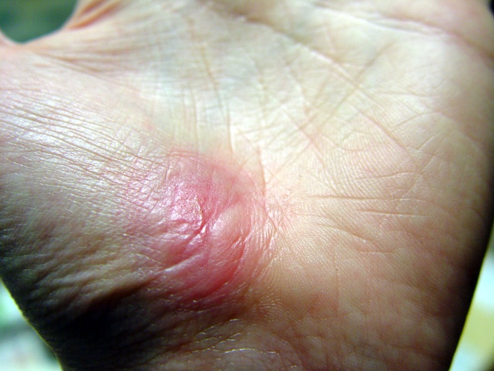
There are several types of bacteria that can infect open wounds and cause a rare condition called necrotizing fasciitis. These bacteria do not merely damage the surface of the skin – they release toxins that destroy the underlying tissue, including muscles, nerves and blood vessels. Once the bacteria reach the bloodstream, they gain ready access to additional tissues and organ systems. If left untreated, necrotizing fasciitis can be fatal, sometimes within 48 hours.

The bacterial species group A Streptococcus, or group A strep, is the most common culprit behind necrotizing fasciitis. But the CDC’s latest warning points to an additional suspect, a type of bacteria called Vibrio vulnificus. There are only 150 to 200 cases of Vibrio vulnificus in the U.S. each year, but the mortality rate is high, with 1 in 5 people succumbing to the infection.

How do you catch flesh-eating bacteria?

Vibrio vulnificus primarily lives in warm seawater but can also be found in brackish water – areas where the ocean mixes with freshwater. Most infections in the U.S. occur in the warmer months, between May and October. People who swim, fish or wade in these bodies of water can contract the bacteria through an open wound or sore.

Vibrio vulnificus can also get into seafood harvested from these waters, especially shellfish like oysters. Eating such foods raw or undercooked can lead to food poisoning, and handling them while having an open wound can provide an entry point for the bacteria to cause necrotizing fasciitis. In the U.S., Vibrio vulnificus is a leading cause of seafood-associated fatality.

Why are flesh-eating bacteria infections rising?

Vibrio vulnificus is found in warm coastal waters around the world. In the U.S., this includes southern Gulf Coast states. But rising ocean temperatures due to global warming are creating new habitats for this type of bacteria, which can now be found along the East Coast as far north as New York and Connecticut. A recent study noted that Vibrio vulnificus wound infections increased eightfold between 1988 and 2018 in the eastern U.S.

Climate change is also fueling stronger hurricanes and storm surges, which have been associated with spikes in flesh-eating bacteria infection cases.

Aside from increasing water temperatures, the number of people who are most vulnerable to severe infection, including those with diabetes and those taking medications that suppress immunity, is on the rise.

What are symptoms of necrotizing fasciitis? How is it treated?

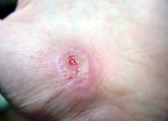
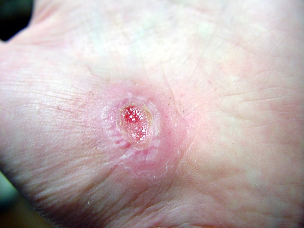
Early symptoms of an infected wound include fever, redness, intense pain or swelling at the site of injury. If you have these symptoms, seek medical attention without delay. Necrotizing fasciitis can progress quickly, producing ulcers, blisters, skin discoloration and pus.

Treating flesh-eating bacteria is a race against time. Clinicians administer antibiotics directly into the bloodstream to kill the bacteria. In many cases, damaged tissue needs to be surgically removed to stop the rapid spread of the infection. This sometimes results in amputation of affected limbs.

Researchers are concerned that an increasing number of cases are becoming impossible to treat because Vibrio vulnificus has evolved resistance to certain antibiotics.

How do I protect myself?

The CDC offers several recommendations to help prevent infection.

People who have a fresh cut, including a new piercing or tattoo, are advised to stay out of water that could be home to Vibrio vulnificus. Otherwise, the wound should be completely covered with a waterproof bandage.

People with an open wound should also avoid handling raw seafood or fish. Wounds that occur while fishing, preparing seafood or swimming should be washed immediately and thoroughly with soap and water.

Anyone can contract necrotizing fasciitis, but people with weakened immune systems are most susceptible to severe disease. This includes people taking immunosuppressive medications or those who have pre-existing conditions such as liver disease, cancer, HIV or diabetes.

It is important to bear in mind that necrotizing fasciitis presently remains very rare. But given its severity, it is beneficial to stay informed.The Conversation


Bill Sullivan, Professor of Pharmacology & Toxicology, Indiana University

This article is republished from The Conversation under a Creative Commons license. Read the original article.

Read More

FDA approves wound retraction system that fights surgical site infections

The FDA has approved a wound retraction system created by Prescient Surgical. The company’s CleanCision wound retraction and protection system is a sterile, single-use irrigating wound device that integrates surgical retraction, wound barrier protection, and fluid delivery and removal to protect against surgical site infections (SSIs).

Read more here.

Read More

Omentum flap as a salvage procedure in deep sternal wound infection

omentum flap procedure deep sternal wound infection wca

Introduction: Deep sternal wound infections (DSWIs) are rare but devastating complication after median sternotomy following cardiac surgery. Especially in the presence of artificial material or inadequate preliminary muscle flaps, the pedicled omentum flap is due to its immunological properties, the predetermined flap in salvage procedures. (more…)

Read More

Preventing Infections in Patients with Wounds eBook

sponsored by Angelini Pharma, Inc.

> Fill out this form to receive your free ebook

|sponsored by Angelini Pharma, Inc.|


by downloading this ebook you are opting in to receiving information from Angelini. 

Read More

Antibiotic use in pressure injury infections

antibiotic overuse pressure injury infection

Antibiotic overuse contributes to the problems of antibiotic resistance and healthcare acquired infections, such as Clostridium difficile. Antibiotic stewardship programs improve patient outcomes, reduce antimicrobial resistance, and save money. These programs are designed to ensure patients receive the right antibiotic, at the right dose, at the right time, and for the right duration. (more…)

Read More

Peptide nanofibers keep infections at bay

Researchers have developed a new infection-blocking material made of peptide-containing nanofibers that works against antibiotic-resistant bacteria and could one day be incorporated into wound dressings (ACS Infect. Dis. 2017, DOI: 10.1021/acsinfec​dis.6b00173).

The approach targets bacterial quorum sensing—a mode of chemical communication used by bacteria to detect other bacteria. When they sense that enough of their kind are present, they can mount an infectious attack.

Read more at Chemical and Engineering News

Read More

Wise use of antibiotics in patients with wound infections

Antibiotic resistance is a pressing public health threat not only in the United States, but worldwide. According to the World Health Organization (WHO), it is one of the major threats to human health.

Despite these concerns, antibiotics continue to be widely used—and overused. In long-term care, for instance, antibiotics are the most frequently prescribed medications, with as many as 70% of residents receiving one or more courses per year. And antibiotics are consistently ordered for suspected pressure ulcer infections.

Here is what clinicians who care for patients with wounds can do to help reduce antibiotic resistance. (more…)

Read More

Clinical Notes: Aspirin, Skin Infections, NPWT surgical incisions

Aspirin inhibits wound healing

A study in the Journal of Experimental Medicine describes how aspirin inhibits wound healing and paves the way for the development of new drugs to promote healing.

The authors of “12-hydroxyheptadecatrienoic (12-HHT) acid promotes epidermal wound healing by accelerating keratinocyte migration via the BLT2 receptor” report that aspirin reduced 12-HHT production, which resulted in delayed wound closure in mice. However, a synthetic leukotriene B4 receptor 2 (BLT2) agonist increased the speed of wound closure in cultured cells and in diabetic mice. (more…)

Read More

Why do older people heal more slowly?

After a few days, the regenerative phase will be well at work closing the wound. Jpbarrass via Wikimedia Commons

By Matthew Steinhauser, University of Pittsburgh

I recently visited an 83-year-old patient in the hospital after EMTs rushed her to the ER with an infected leg wound. Her ordeal started inconspicuously when she bumped into the sharp edge of a table and developed a small cut. The patient’s wound didn’t close, but she ignored it until she woke up in pain one morning two weeks after first injuring her leg. Her daughter called 911 after noticing angry, red skin discoloration and pus – both signs of an infection. Our medical team treated her with IV antibiotics and cleared up the infection, but the wound did not fully close until at least a month later, well after she was discharged from the hospital.

How different the story is when children get a cut. They may scream initially, but within days, the scab falls off, revealing new skin. Why was healing so delayed in my 83-year-old patient compared to a healthy child?

The answer is age. Decades of life slow down healing for most tissues, and wounds in skin can offer a window into why this slowdown occurs.

Three stages of wound healing

I am physician who studies how aging predisposes patients to diseases like diabetes and whether behavioral changes such as intermittent fasting may slow down aging. In order to understand why the skin wound in my older patient healed so slowly, it is important to first understand how wounds heal under the ideal conditions of youth.

The wound healing process is classically categorized into three stages.

A fresh scrape on a palm right after it occured.


Right after a wound occurs, the inflammatory response begins.
Jpbarrass via Wikimedia Commons

The first stage is inflammation, essentially the body’s attempt to clean the wound. During the inflammatory phase, immune cells called phagocytes move into the wound, kill any contaminating bacteria, and ingest and dispose of dead cells and debris.

An image of a hand with a partially healed scrape.
After a few days, the regenerative phase will be well at work closing the wound.
Jpbarrass via Wikimedia Commons

Inflammation sets the stage for the regenerative phase, where several processes work in concert to regrow damaged skin. Replacement skin cells are born when cells at the edge of the wound divide, while fibroblast cells lay down a supportive scaffolding called the extracellular matrix. This holds the new cells together. Any damaged supporting structures of the skin, such as the blood vessels that supply critical oxygen and nutrients, also need to regrow. The second stage effectively closes the wound and restores a protective barrier against bacteria.

A cut on someones hand mostly healed over and scarring.
Once the wound is fully closed, the remodeling phase will rebuild the tissue in a stronger way.
Jpbarrass via Wikimedia Commons

The regenerative phase is a relatively quick, but tenuous fix – new skin is fragile. The final remodeling phase plays out over a couple of years as the new skin is progressively strengthened by several parallel processes. The extracellular matrix, which was initially laid down in a haphazard fashion, is broken down and replaced in a more durable way. Any residual cells from prior phases that are no longer needed – such as immune cells or fibroblasts – become inactive or die. In addition to strengthening the new skin, these collective actions also account for the tendency of scars to visibly fade with time.

Diseases disrupt the healing process

One major way aging can derail the orderly and efficient progression through the stages of healing is through the health problems that stem from diseases of old age.

Diabetes is one example of a disease that is strongly associated with older age. One of the many ways that diabetes negatively affects healing is by causing blood vessels to narrow. As a consequence of inadequate circulation, crucial nutrients and oxygen do not reach the wound in sufficient quantities to fuel the second regenerative phase.

Diabetes is just one of many age-related diseases that disrupts normal processes in the body such as wound healing.

Cells age too

Aside from the negative impacts of age-associated diseases, cells themselves age. In an extreme sign of aging called cellular senescence, cells permanently lose the ability to divide. Senescent cells accumulate in skin and many other organs as people age and cause a host of problems.

When cells divide more slowly – or when they stop dividing altogether due to senescence – skin becomes thinner. The replacement of fat cells, which form a cushioning layer under the skin, also declines with age. The skin of older patients is therefore more prone to injury in the first place.

Once an older person’s skin is injured, the skin has a harder time healing properly as well. Aging and senescent immune cells cannot defend against bacteria, and the risk of serious skin infection rises. Then in the regenerative stage, slow rates of cell division translate into slow skin regrowth. My patient exhibited all of these negative effects of age – her thin, almost translucent skin ruptured from a minor bump, became infected and took nearly two months to fully regrow.

But senescent cells are more than just dysfunctional bystanders. For reasons that are not yet fully understood, senescent cells release toxic byproducts that damage surrounding tissue and drive inflammation – even when there’s no bacterial threat present. Some of these byproducts can even accelerate senescence in neighboring cells. This suggests that intrinsic aging of cells is in essence contagious and senescent cells actively fuel an uncontrolled cycle of inflammation and tissue damage that further impedes successful regeneration and healing.

A whole body problem

As the most outwardly visible tissue of the body, the skin provides a window into why people heal more slowly with age, but all tissues can be injured and are susceptible to the effects of aging. Injuries may be small, repetitive and build up over time – like the effect of smoking on the lungs. Or they may be discrete and dramatic – such as the death of heart cells with a heart attack. Different tissues may heal in different ways. Yet all tissues share a sensitivity to the repercussions of an aging immune system and a decline in the ability to regrow dead or damaged cells.

Understanding why healing slows down with age is important, but my patient asked a very practical question that physicians often face in one form or another: “Doctor, what can you do for me?”

Unfortunately, current treatment of wounds is fairly old-fashioned and often ineffective. Some of the options available include wound dressing changes, antibiotics when the wound is infected or treatment in a high oxygen chamber when circulation is bad due to diabetes.

There is hope, though, that medicine can do better and that progress in understanding the aging process will lead to new therapies. Neutralizing senescent cells in mice, for example, improves a variety of age-associated diseases. While it is way too early to say that researchers have discovered the fountain of youth, I am optimistic for a future when physicians will bend the aging curve and make skin and other organs heal faster and better.The Conversation


Matthew Steinhauser, Associate Professor of Medicine, University of Pittsburgh

This article is republished from The Conversation under a Creative Commons license. Read the original article.

Read More

A young Black scientist discovered a pivotal leprosy treatment in the 1920s − but an older colleague took the credit

Dr. Isabel Kerr, a European missionary, administering to a patient a chaulmoogra oil treatment in 1915, prior to the invention of the Ball Method. George McGlashan Kerr, CC BY

By Mark M. Lambert, Des Moines University

Hansen’s disease, also called leprosy, is treatable today – and that’s partly thanks to a curious tree and the work of a pioneering young scientist in the 1920s. Centuries prior to her discovery, sufferers had no remedy for leprosy’s debilitating symptoms or its social stigma.

This young scientist, Alice Ball, laid fundamental groundwork for the first effective leprosy treatment globally. But her legacy still prompts conversations about the marginalization of women and people of color in science today.

As a bioethicist and historian of medicine, I’ve studied Ball’s contributions to medicine, and I’m pleased to see her receive increasing recognition for her work, especially on a disease that remains stigmatized.

Who was Alice Ball?

Alice Augusta Ball, born in Seattle, Washington, in 1892, became the first woman and first African American to earn a master’s degree in science from the College of Hawaii in 1915, after completing her studies in pharmaceutical chemistry the year prior.

After she finished her master’s degree, the college hired her as a research chemist and instructor, and she became the first African American with that title in the chemistry department.

Impressed by her master’s thesis on the chemistry of the kava plant, Dr. Harry Hollmann with the Leprosy Investigation Station of the U.S. Public Health Service in Hawaii recruited Ball. At the time, leprosy was a major public health issue in Hawaii.

Doctors now understand that leprosy, also called Hansen’s disease, is minimally contagious. But in 1865, the fear and stigma associated with leprosy led authorities in Hawaii to implement a mandatory segregation policy, which ultimately isolated those with the disease on a remote peninsula on the island of Molokai. In 1910, over 600 leprosy sufferers were living in Molokai.

This policy overwhelmingly affected Native Hawaiians, who accounted for over 90% of all those exiled to Molokai.

The significance of chaulmoogra oil

Doctors had attempted to use nearly every remedy imaginable to treat leprosy, even experimenting with dangerous substances such as arsenic and strychnine. But the lone consistently effective treatment was chaulmoogra oil.

Chaulmoogra oil is derived from the seeds of the chaulmoogra tree. Health practitioners in India and Burma had been using this oil for centuries as a treatment for various skin diseases. But there were limitations with the treatment, and it had only marginal effects on leprosy.

The oil is very thick and sticky, which makes it hard to rub into the skin. The drug is also notoriously bitter, and patients who ingested it would often start vomiting. Some physicians experimented with injections of the oil, but this produced painful pustules.

Dr. Isabel Kerr, a European missionary, administering to a patient a chaulmoogra oil treatment in 1915, prior to the invention of the Ball Method. George McGlashan Kerr, CC BY

The Ball Method

If researchers could harness chaulmoogra’s curative potential without the nasty side effects, the tree’s seeds could revolutionize leprosy treatment. So, Hollmann turned to Ball. In a 1922 article, Hollmann documents how the 23-year-old Ball discovered how to chemically adapt chaulmoogra into an injection that had none of the side effects.

The Ball Method, as Hollmann called her discovery, transformed chaulmoogra oil into the most effective treatment for leprosy until the introduction of sulfones in the late 1940s.

In 1920, the Ball Method successfully treated 78 patients in Honolulu. A year later, it treated 94 more, with the Public Health Service noting that the morale of all the patients drastically improved. For the first time, there was hope for a cure.

Tragically, Ball did not have the opportunity to revel in this achievement, as she passed away within a year at only 24, likely from exposure to chlorine gas in the lab.

Ball’s legacy, lost and found

Ball’s death meant she didn’t have the opportunity to publish her research. Arthur Dean, chair of the College of Hawaii’s chemistry department, took over the project.

Dean mass-produced the treatment and published a series of articles on chaulmoogra oil. He renamed Ball’s method the “Dean Method,” and he never credited Ball for her work.

Ball’s other colleagues did attempt to protect Ball’s legacy. A 1920 article in the Journal of the American Medical Association praises the Ball Method, while Hollmann clearly credits Ball in his own 1922 article.

Ball is described at length in a 1922 article in volume 15, issue 5, of Current History, an academic publication on international affairs. That feature is excerpted in a June 1941 issue of Carter G. Woodson’s “Negro History Bulletin,” referring to Ball’s achievement and untimely death.

Joseph Dutton, a well-regarded religious volunteer at the leprosy settlements on Molokai, further referenced Ball’s work in a 1932 memoir broadly published for a popular audience.

Historians such as Paul Wermager later prompted a modern reckoning with Ball’s poor treatment by Dean and others, ensuring that Ball received proper credit for her work. Following Wermager’s and others’ work, the University of Hawaii honored Ball in 2000 with a bronze plaque, affixed to the last remaining chaulmoogra tree on campus.

In 2019, the London School of Hygiene and Tropical Medicine added Ball’s name to the outside of its building. Ball’s story was even featured in a 2020 short film, “The Ball Method.”

The Ball Method represents both a scientific achievement and a history of marginalization. A young woman of color pioneered a medical treatment for a highly stigmatizing disease that disproportionately affected an already disenfranchised Indigenous population.

In 2022, then-Gov. David Ige declared Feb. 28 Alice Augusta Ball Day in Hawaii. It was only fitting that the ceremony took place on the Mānoa campus in the shade of the chaulmoogra tree.The Conversation


Mark M. Lambert, Assistant Professor of Behavioral Medicine, Medical Humanities, and Bioethics, Des Moines University

This article is republished from The Conversation under a Creative Commons license. Read the original article.

Read More
1 2 3 15