We all lead busy lives, with demanding work schedules and home responsibilities that can thwart our best intentions. Although we know it’s our responsibility to stay abreast of changes in our field, we may feel overwhelmed when we try to make that happen.
Keeping clinicians up-to-date on clinical knowledge is one of the main goals of the Wild On Wounds (WOW) conference, held each September in Las Vegas. Each year, I present the opening session of this conference, called “The Buzz Report,” which focuses on the latest-breaking wound care news—what’s new, what’s now, what’s coming up. I discuss innovative new products, practice guidelines, resources, and tools from the last 12 months in skin, wound, and ostomy management. This article highlights the hottest topics from my 2015 Buzz Report.
Guidelines buzz
Although not new in 2015, “Prevention and Treatment of Pressure Ulcers: Clinical Practice Guideline” from the National Pressure
Ulcer Advisory Panel (NPUAP), European Pressure Ulcer Advisory Panel, and Pan Pacific Pressure Injury Alliance is still a buzzing topic. The guideline was released in September 2014, and many facilities and clinicians are still busy trying to incorporate it into their protocols. This can be an arduous task, given the more than 575 specific recommendations. However, the quick-pick system using “thumbs up” and “thumbs down” icons next to each recommendation helps users separate the should do’s from the don’t do’s.
The American College of Physicians released two pressure ulcer guidelines in March 2015. “Treatment of Pressure Ulcers: A Clinical Practice Guideline” and “Risk Assessment and Prevention of Pressure Ulcers” are based on a systematic evidence review and focus on specific aspects of care. Each guideline has just three recommendations.
Although not a guideline per say, the evidence-based consensus document “The Management of Diabetic Foot Ulcers (DFUs) Through Optimal Off-loading” published in the Journal of the American Podiatric Medical Association includes eight specific consensus statements. Here are two of the most notable:
• Consensus statement #4: Total contact casting is the preferred method for off-loading plantar DFUs, as it has most consistently demonstrated the best healing outcomes and is a cost-effective treatment.
• Consensus statement #5: There currently exists a gap between the evidence supporting the efficacy of DFU off-loading and what is performed in clinical practice.
Literature buzz
Thousands of wound and ostomy articles are published each year. Below are just a few of the articles that I believe will have a significant impact at the bedside.
“What is the healing time of Stage II pressure ulcers? Findings from a secondary analysis,” in Advances in Skin & Wound Care Journal, describes data collected from a multicenter randomized clinical trial. The authors conclude that achieving complete re-epithelialization in stage 2 pressure ulcers takes approximately 23 days and that on average, small ulcers heal 12 days faster than those with a surface of 3.1 cm2 or greater.
NPUAP released two key papers in 2015.
• “Hand check method: Is it an effective method to monitor for bottoming out?” reviewed the science behind the clinical practice of hand checks for bottoming out on a support surface. NPUAP’s position statement supports use of hand checks with air mattress overlays and chair cushions only. NPUAP stated more research is needed to develop acceptable ways to evaluate the performance of mattress replacements and integrated bed systems; until such time, clinicians should follow the manufacturer’s recommendation and not perform hand checks.
• The white paper “Do lift slings significantly change the efficacy of therapeutic support surfaces?” is designed to increase clinicians’ critical thinking when using lift slings in combination with therapeutic support surfaces. NPUAP recommends clinicians choose a combination of support surface and sling that meets the patient’s needs while focusing on the risks and benefits of leaving a sling beneath a patient.
A 2015 review and analysis of literature on friction and pressure ulcers in the Journal of Wound Ostomy Continence Nursing explained that friction alone doesn’t directly cause pressure ulcers, and cautioned against categorizing friction wounds as pressure ulcers. “Friction-induced skin injuries—are they pressure ulcers? An updated NPUAP white paper” explains that friction can result in shear forces that may lead to a pressure ulcer; however, without shear, friction alone doesn’t lead to pressure ulcers.
Ulcers from sickle cell disease
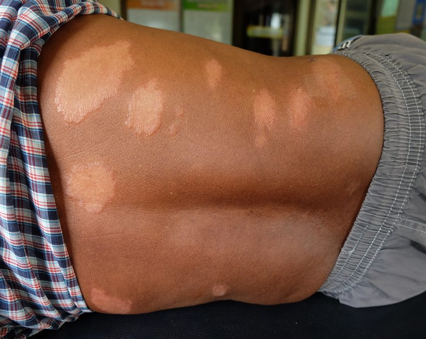
About 1% to 3% of the U.S. population lives with sickle cell disease (SCD). From 25% to 75% of these people also experience leg ulcers. “Sickle cell disease & wound care: Lower extremity ulcers in ‘crisis,’” published in Today’s Wound Clinic, identified key diagnostic characteristics and treatment protocols to consider. The underlying cause of SCD ulcers remains unknown. Most begin spontaneously or from trauma as small scabbed areas over the medial or lateral malleoli. Scabs progress to round, punched-out lesions with raised margins, deep bases, and necrotic slough, with surrounding brown hyperpigmentation and scaling. Patients typically complain of extreme tenderness or pain at the ulcer site.
Treatment aims to manage SCD and associated anemia and control pain. Local wound care involves moist wound healing, bacteria control, protection from trauma, loose-fitting clothing around the ankles to avoid friction, and pressure dressings, such as an Unna’s boot. In many cases, sharp debridement can’t be done because of intolerable pain. A good alternative is biological debridement.
Infrared skin thermometry
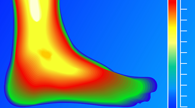
All objects at temperatures above absolute zero release infrared radiation. Heat from wound inflammation, fever, and infection is a form of infrared radiation. By using a noncontact infrared thermometer to monitor wounds and surrounding tissue, clinicians can identify signs of deep inflammation, infection, or trauma that may be invisible on the surface. “Infrared skin thermometry: An underutilized cost-effective tool for routine wound care practice and patient high-risk diabetic foot self-monitoring,” published in Advances in Wound Care, found wounds with an elevated temperature measured with infrared thermometry were eight times more likely to be diagnosed with deep infection. A temperature elevation over the same spot on the other foot in a patient with diabetes without a foot ulcer may indicate an acute Charcot foot. In addition, limb ischemia results in lower regional, local, and side-to-side variability in temperatures. Using an infrared thermometer, clinicians can identify unequal vascular supply by measuring temperatures proximal and distal to the wound. Commercially available, inexpensive, noncontact infrared thermometers can detect localized increases in skin surface temperature comparable to scientific grade instruments.
Noncontact infrared thermometry also can be used to assess the skin for pressure ulcers, such as deep-tissue injury, dark skin tones, and circulatory status around the wound. I believe all wound care practitioners should have a noncontact infrared skin thermometer on their tool belt. For examples of these thermometers, visit http://goo.gl/6wN5eJ.
Product buzz
Debrisoft® is a ground-breaking active debridement system from Lohmann & Rauscher that mechanically debrides and cleans wounds by rapidly removing debris, necrotic material, slough, exudate, and hyperkeratotic tissue. The dressing is made of soft, angled polyester fibers that loosen debris while protecting intact granulation tissue and epithelial cells. To use, moisten with tap water or saline solution. Then, using light pressure and a circular motion, gently rub the wound or skin with the soft, fleecy side of the dressing. You can use Debrisoft each time you change the wound dressing.
A similar product, DebriMitt™ from Crawford Healthcare, is designed as a single-use mitt with a finger pouch. It gently removes nonviable tissue, hyperkeratotic skin, and debris and can disrupt biofilms in the wound base.
A natural approach to wound debridement can be achieved with the new BioMonde BioBag®, which contains disinfected larvae of Lucilia sericata (maggots) in a sealed sterile polyester net bag. The bag is placed directly onto the wound bed; larvae remain sealed within the dressing for the full 4-day treatment. The BioBag allows larvae to pass secretions through the pores of the polyester containment net, dissolving and physically removing devitalized tissue and bacteria from the wound without removing healthy and viable tissue. All wound-cleaning benefits of larval therapy remain in the BioBag without fear of larvae wandering from the treatment area.
Helix3 CM™ and Helix3 CP™ are new collagen wound dressings from Amerx. Helix3 CM is a bioactive collagen matrix dressing composed of 100% type 1 bovine native collagen formulated in a highly absorptive porous collagen sheet. Helix3 CP is 100% type 1 bovine nonhydrolyzed collagen powder. Because these products aren’t hydrolyzed, they contain 10 times more nondenatured, native triple-helix structured collagen than similar products.
For the latest bedding fabrics that reduce shear and friction, see New bedding fabrics.
Note: Watch for part 2 of the Buzz Report in the March-April issue.
Selected references
Brienza D, Antokal S, Herbe L, et al. Friction-induced skin injuries: Are they pressure ulcers? An updated NPUAP white paper. J Wound Ostomy Continence Nurs. 2015;42(1):62-4
Brienza D, Deppisch M, Gillespie C. Do lift slings significantly change the efficacy of therapeutic support surfaces? A National Pressure Ulcer Advisory Panel White Paper. March 2015. http://goo.gl/nocsIj
Call E, Deppisch M, Jordan R, et al. Hand check method: Is it an effective method to monitor for bottoming out? A National Pressure Ulcer Advisory Panel Position Statement. June 2015. http://goo.gl/k0U4OL
National Pressure Ulcer Advisory Panel, European Pressure Ulcer Advisory Panel, and Pan Pacific Pressure Injury Alliance. Prevention and Treatment of Pressure Ulcers: Quick Reference Guide. Emily Haesler, ed. Perth, Australia: Cambridge Media; 2014. http://goo.gl/5IkUVG
Palese A, Luisa S, Ilenia P, et al; PARI-ETLD Group. What is the healing time of Stage II pressure ulcers? Findings from a secondary analysis. Adv Skin Wound Care. 2015;28(2):69-75.
Penne JR, Goodman BM, Chen IA. Sickle cell disease & wound care: lower extremity ulcers in “crisis.” Today’s Wound Clinic. 2015;9(3). http://goo.gl/nfEk68
Qaseem A, Humphrey LL, Forciea MA, et al; Clinical Guidelines Committee of the American College of Physicians. Treatment of pressure ulcers: a clinical practice guideline from the American College of Physicians. Ann Intern Med. 2015;162(5):370-9.
Qaseem A, Mir TP, Starkey M, et al; Clinical Guidelines Committee of the American College of Physicians. Risk assessment and prevention of pressure ulcers: a clinical practice guideline from the American College of Physicians. Ann Intern Med. 2015;162(5):359-69.
Sibbald RG, Mufti A, Armstrong DG. Infrared skin thermometry: an underutilized cost-effective tool for routine wound care practice and patient high-risk diabetic foot self-monitoring. Adv Skin Wound Care. 2015;28(1):37-44.
Snyder RJ, Frykberg RG, Rogers LC, et al. The management of diabetic foot ulcers through optimal off-loading: building consensus guidelines and practical recommendations to improve outcomes. J Am Podiatr Med Assoc. 2014;104(6):555-67.
Online Resources
O. therapeuticbedding.com/more_woundcare
Donna Sardina is editor-in-chief of Wound Care Advisor and cofounder of the Wound Care Education Institute in Plainfield, Illinois.
DISCLAIMER: All clinical recommendations are intended to assist with determining the appropriate wound therapy for the patient. Responsibility for final decisions and actions related to care of specific patients shall remain the obligation of the institution, its staff, and the patients’ attending physicians. Nothing in this information shall be deemed to constitute the providing of medical care or the diagnosis of any medical condition. Individuals should contact their healthcare providers for medical-related information.