Legal Matters

What caused Mr. M’s pressure ulcer?

A court case answers the question as to whether a pressure ulcer was preventable

By Nancy J. Brent, MS, RN, JD

Pressure ulcers are a major health risk for every adult patient. Risk factors include sepsis, hypotension, and age 70 or older. These risk factors became all too real when Mr. M developed pressure ulcers after being admitted to a Texas hospital.

Background

Mr. M, age 81, presented at a medical center’s emergency department on January 2 complaining of abdominal pain. After undergoing an assessment, he was diagnosed with gallstones and admitted to the hospital. The next day, he had gallbladder surgery. He subsequently developed a bowel obstruction and had to undergo two more surgeries for this condition over the next 10 days.
On January 13, he was transferred to the intensive care unit (ICU) because of multiple serious medical conditions, including respiratory distress syndrome (necessitating ventilatory support), septic shock, a “blood infection” that caused his blood pressure to drop, and multiorgan failure. His primary physician discontinued tube feedings out of concern they might exacerbate his renal failure; he wrote a do-not-resuscitate order and ordered sedation.
Mr. M was unable to turn or position himself in any way. While in the ICU, he developed a “skin tear” on the tailbone (coccyx) that progressed to a serious pressure ulcer. On February 6, his condition improved enough to allow his transfer to a rehabilitation hospital, where he developed pressure ulcers on his heels. He was transferred to another hospital; the ulcer on his coccyx healed by August. He remained in that hospital for 1 year before being discharged home.
Despite healing of the pressure ulcer on his coccyx, the wound area remained hard and painful, and Mr. M experienced “daily discomfort” there. Also, he was unable to do many of the things he’d been able to do before his hospitalization.

Mr. M files a medical malpractice suit

Mr. M sued the medical center, alleging the hospital was negligent by failing to prevent the pressure ulcer from forming through the use of known “pressure relief” methods, and that the hospital failed to provide proper care and treatment of the wound once it was discovered.
At trial, the medical center lawyers argued that Mr. M’s grave condition caused the pressure ulcer to develop. The jury returned a verdict for Mr. M, finding that the medical center’s negligence proximately caused the injuries he sustained. It awarded him $35,000 for medical expenses; $135,000 for past physical pain and mental anguish; $25,000 for future physical pain and mental anguish; $25,000 for past physical impairment; and $25,000 for future physical impairment. The medical center appealed the decision.

Medical center appeals the verdict

Several issues were raised by the medical center on appeal. Of particular interest to nurses and wound care practitioners was the “cause in fact” or the “proximate cause” of Mr. M’s pressure ulcer on the coccyx. Because an expert witness must establish proximate cause based on a reasonable degree of medical certainty, Mr. M’s case became a battle of the experts regarding the care he received, or lack of care, relative to development of the pressure ulcer.

Expert witness testimony for Mr. M

The first nurse expert to testify was Mr. M’s highly qualified expert. She testified about the various acceptable ways to provide pressure relief, including turning the patient or, if the patient can’t be turned, repositioning. The latter requires use of foam wedges or pillows to elevate a particular body part. The nurse expert testified that if a patient can’t be turned or repositioned, that fact must be documented along with the reason for inability to carry out this nursing care.
Proper assessment of the pressure ulcer is required so that other team members can “see” the wound; the clinician who assesses the wound should draw a picture of exactly what he or she saw when documenting the note in the patient’s chart. The nurse expert testified that the assessment should include the color, duration, and depth of the pressure ulcer; presence or absence of infection; and whether the tissue was dead or perfused.
After reviewing the medical center’s policies and protocols on pressure relief, which required nurses to provide pressure relief every 2 hours, and the depositions of the nurses who’d cared for Mr. M, the nurse expert testified there was no documentation showing Mr. M received any pressure relief from January 13 to January 16. She said she could only conclude that the nurses failed to turn or reposition him during those days. The only notation made about his skin condition was when nurses discovered the “skin tear” on January 14. After this discovery, the physician wasn’t notified of it until January 19. On that date, the physician ordered a wound care consult, but the actual consultation didn’t occur until 3 days later. Even with the wound consultant’s specific, written orders to care for the wound, only one notation existed showing that the orders were followed. Also, the wound care orders weren’t entered into Mr. M’s care plan until January 28. Additionally, in their depositions, the nurses caring for Mr. M couldn’t recall changing the dressing as ordered.
Therefore, in the nurse expert’s opinion, the pressure ulcer on Mr. M’s coccyx was caused directly by failure of the ICU nurses to provide pressure relief from January 14 to January 16 and that providing the wound care that was ordered would have prevented the ulcer from getting worse and would have healed the ulcer.
Although a physician serving as a second expert for Mr. M also testified that pressure relief should have been provided, he couldn’t say that development of the pressure ulcer was unpreventable.

Expert witness testimony for the medical center

Not surprisingly, the medical center’s expert witnesses, two of whom were physicians, testified that because of Mr. M’s general medical condition, he would have developed the pressure ulcer even if hospital policies and protocols had been followed. The hospital’s nurse expert witness stated that Mr. M’s pressure ulcer was not preventable because of his medical condition, regardless of whether or not he was turned. In her opinion, the active range of motion his nurses put him through was enough to reperfuse the area.

Appellate court’s decision

The appellate court upheld the trial court jury’s verdict, stating that evidence presented at the trial was legally and factually sufficient to support that verdict.

Take-away points

Mr. M’s case undoubtedly was complicated by his age and general medical condition, as well as disagreement among expert witnesses as to the cause of the pressure ulcer on his coccyx. Even so, the appellate court held that the evidence at trial (specifically that presented by Mr. M’s nurse expert witness) was sufficient legally and factually to support the verdict in favor of Mr. M.
This case illustrates many areas of importance for nurses in terms of formation and care of pressure ulcers. They include the following:
• Risk factors supporting potential formation of pressure ulcers can’t be overlooked or underestimated by nursing staff.
• A plan to prevent pressure ulcers should be initiated on admission for every patient who is immobile or has other risk factors for pressure ulcers.
• Documentation of every aspect of nursing care that’s initiated and continued to prevent pressure ulcers from forming must be carried out as ordered and pursuant to hospital policy and protocol.
• Care plans, communications with other health team members, and carrying out of orders must be done as soon as possible.
• Assessment and documentation of pressure ulcers should include enough detail so other health team members can visualize what the nurse entering the documentation has seen.
• The nurse should assess and stage the pressure ulcer at each dressing change.
• One’s expert witness must be credentialed, educated, and experienced in would care prevention and treatment, because his or her testimony can win or lose a case.

Nursing remains at the forefront of protecting and safeguarding patients from pressure ulcers. Although not every ulcer can be prevented, the goal is to prevent as many ulcers as possible. If a pressure ulcer does occur, caregivers’ essential focus must be on healing or preventing further deterioration and infection.

Selected references
Columbia Medical Center Subsidiary, L.P., d/b/a/
North Central Medical Center, Appellant, v. John Meier, Appellee. 198 S.W. 3d 408 (Ct. Appeals 2006).

Lyder CH, Ayello EA. Pressure ulcers: A Patient Safety Issue. In: Hughes RG, ed. Patient Safety and Quality: An Evidence-Based Handbook For Nurses. Rockville, MD: Agency For Healthcare Research and Quality. April 2008. www.ncbi.nlm.nih.gov/books/
NBK2650/
. Accessed November 1, 2012.

Nancy J. Brent is an attorney in Wilmette, Illinois. The information in this article is for educational purposes only and doesn’t constitute legal advice.

Read More

Clinician Resources

There’s an app for that! Here are a variety of medical apps that you might want to try. You can download them in the iTunes store, and the basic service is free.

Medscape

More than 1.4 million healthcare professionals use this app from WebMD, which includes:
• medical news
• clinical reference information, such as drugs and diseases
• medical calculators (not available for iPad).
The app is available for Android, iPad, and iPhone/iPod touch devices.

http://medscape.com

Pressure Wound Analyzer

Use this app to take a photo of a wound. The app segments the image into red, yellow, and black to help with ulcer classification. You can also use the app to track changes in the wound over time. The app is available for iPad and iPhone/iPod touch devices. Note: This is free for a limited time.

http://woundanalyzer.blogspot.com

AHRQ ePSS

The ePSS (Electronic Preventive Services Selector) app allows you to search and browse the U.S. Preventive Services Task Force recommendations on the Web or a mobile device. The app is from the U.S. Department of Health & Human Services and is available for Android, iPad, and iPhone/iPod touch devices.

http://epss.ahrq.gov

UMSkinCheck

This app from the University of Michigan Health System allows users to complete and store photographs of the skin. Features include:
• guidance on performing a skin cancer self-exam and full-body photographic survey
• tracking of detected skin lesions and moles for changes over time
• notifications/reminders to perform self-exams on a routine basis
• storage of photos for baseline comparisons during routine follow-up self-
exams
• informational videos and literature on skin cancer prevention and healthy skin as well as a skin cancer risk calculator function.
The app is available for iPad and iPhone/iPod touch devices.

http://uofmhealth.org/patient%20and%20visitor%20guide/my-skin-check-app

Glucose Buddy—Diabetes Logbook Manager w/syncing, blood pressure, weight tracking

This data storage utility app is perfect for your patients with diabetes who want all their information in one place. Users can manually enter their glucose results, carbohydrate consumption, insulin dosages, and activities, and then view the data in a free glucosebuddy.com online account. Another option is the ability to set reminders for when it’s time to check blood glucose.
The app is available for Android, iPad, and iPhone/iPod touch devices.

http://www.glucosebuddy.com

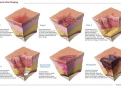
3M Health Care Pressure Ulcer Staging

Use this app to learn more about pressure ulcer staging. It includes information about 3M pressure ulcer products.
The app is available for iPad and iPhone/iPod touch devices.

https://itunes.apple.com/us/app/3m-health-care-pressure-ulcer/id454537520?mt=8

SACS™ Instrument

This evidence-based tool is helpful for assessing and classifying peristomal skin lesions. Click here for more information about the SACS Instrument.
The app is available for iPad and iPhone/iPod touch devices.

http://www.convatec.com/en/cvtus-homeus/cvt-home/0/home/0/393/0/default.html?
hwcr=flex

Read More

Business Consult

­

Feel more relaxed with restorative yoga

By Lisa Marie Bernardo, PhD, MPH, RN, HFI, RYT

Do you experience chronic stress? Is your body stiff and inflexible? Does your mind seem dull and sluggish, your spirit exhausted?
Restorative yoga may help “open” your joints, ease your mind, and revive your spirit. It’s based on the concept that we’re overstimulated and don’t get enough rest. Constant stimulation activates the sympathetic nervous system, overtaxing the fight-or-flight response. The body responds by increasing cortisol and glucose production, which (along with additional unhealthy responses) raises the risk of metabolic syndrome.
Restorative yoga promotes active relaxation, helping to halt the overstimulation cycle. It promotes balance by alternately stimulating and relaxing the body, which is supported in yoga poses with such props as blankets, pillows, yoga mat, and eye covers. Research suggests restorative yoga may ease hot flashes in postmenopausal women and may promote a calm, positive mood in women with ovarian or breast cancer.

Five facets of restorative yoga

Restorative yoga takes a five-faceted approach to relieve the effects of stress. Over time, you’re likely to notice a new awareness of and appreciation for your body, mind, and spirit.
Simply put, networking is an information exchange, a forum for communicating your needs or agenda and, in return, listening and responding to others’ needs or agendas. Good networking requires emotional reciprocity, which means caring about the needs and agendas of the people you network with. Caring about others’ needs is what nurses do, so networking really shouldn’t be that difficult for a nurse.
1. Using props, restorative yoga supports the body in yoga poses, helping muscles and joints release tension and achieve muscular balance.
2. The restorative poses move the spine in all directions—flexion, extension, rotation, and lateral flexion. This enhances spinal flex­-ibility, lubricates vertebrae, and strengthens the deep muscles that stabilize the spine.
3. Inverted poses, in which the feet and legs are elevated, counter the effects of gravity and promote lymph and fluid drainage to the heart.
4. The poses compress and release internal organs, cleansing them while aiding removal of cellular waste and renewing oxygen and nutrients.
5. Finally, the poses balance the body’s male (prana) and female (apana) energies.

Learning the poses

To learn the poses, consider taking a restorative yoga class. (See Finding an instructor.) Beforehand, make sure to tell the instructor about any special health concerns you have, so the instructor can modify the poses for you. Expect to bring your own blankets, pillows, eye covers, and yoga mat. The class will last from 60 to 90 minutes.
Restorative yoga typically doesn’t involve active (hatha) yoga poses, although it may include stretching poses to warm muscles and joints before the restorative poses begin. The instructor will help you use your props to make the poses right for you, and will direct you into a pose using them. Expect to stay in the pose for 5 to 10 minutes. The instructor will guide you by helping you focus on your breath and turn your attention inward. If your mind wanders and your body stays active, accept this reaction and don’t judge yourself. Over time, you’ll learn to use your breath to release tension and to focus and calm your mind.
After you hold the pose for the required duration, the instructor will help you into the next one. Generally, the class is near-silent, with minimal talking; the lights are low and music may play.
W­hen the class ends, you may feel more relaxed and in touch with yourself. If you feel restless and jittery instead, accept your reaction. Don’t judge yourself. Try again. Give yourself the opportunity to experience something different.
Restorative yoga is just one method to renew and reconnect with your inner being. Only you can know if it’s right for you. n

Selected references
Cohen B, Kanaya A, Macer J, Shen H, Chang A, Grady D. Feasibility and acceptability of restorative yoga for treatment of hot flushes: a pilot trial. Maturitas. 2007;56(2):198-204.
Danhauer SC, Tooze JA, Farmer DF, Campbell CR, McQuellon RP, Barrett R, Miller BE. Restorative yoga for women with ovarian or breast cancer: findings from a pilot study. J Soc Integr Oncol. 2008;6(2):47-58.

Lisa Marie Bernardo is the managing member of The PIlates Centre, LLC, in Hampton Township, Pennsylvania, and adjunct faculty at Carlow University School of Nursing in Pittsburgh, Pennsylvania.

To find a certified yoga instructor in your area, check the Yoga Alliance website at www.yogaalliance.com.  Restorative Yoga Teachers (www.restorativeyogateachers.com) focuses exclusively on restorative yoga. This site is operated by Judith Hansen Lasater, PhD, the leader in restorative yoga practice.

Read More

Clinical Notes

NPUAP releases new position statement on exposed cartilage as Stage IV ulcer

The National Pressure Ulcer Advisory Panel (NPUAP) has released a new position statement, “Pressure ulcers with exposed cartilage are Stage IV pressure ulcers,” which states that pressure ulcers with exposed cartilage should be classified as Stage IV.
NPUAP notes that although the presence of “visible or palpable cartilage at the base of a pressure ulcer” wasn’t included in Stage IV terminology, cartilage “serves the same anatomical function as bone,” so it fits into the current Stage IV definition, “Full thickness tissue loss with exposed bone, tendon or muscle. Slough or eschar may be present on some parts of the wound bed. Often including undermining and tunneling.”

Medicare expenditures for diabetic foot care varies significantly by region

Medicare spending on patients with diabetes who have foot ulcers and lower extremity amputations varies significantly by region, according to a study in Journal of Diabetes and Its Complications, but more spending doesn’t significantly reduce 1-year mortality.
Geographic variation in Medicare spending and mortality for diabetic patients with foot ulcers and amputations” examined data from 682,887 patients with foot ulcers and 151,752 patients with lower extremity amputations.
Macrovascular complications in patients with foot ulcers were associated with higher spending, and these complications in patients with amputations were more common in regions with higher mortality rates.
Rates of hospital admission were associated with higher spending and increased mortality rates for patients with foot ulcers and amputations.
“Geographic variation in Medicare spending and mortality rates for diabetic patients with foot ulcers and amputations is associated with regional differences in the utilization of inpatient services and the prevalence of macrovascular complications,” the study concludes.

Patients who develop pressure ulcers in hospital more likely to die

Medicare patients who develop pressure ulcers in the hospital are more likely to die during the hospital stay, have longer lengths of stay, and to be readmitted within 30 days after discharge, according to a study of 51,842 patients in the Journal of the American Geriatrics Society.
Hospital-acquired pressure ulcers: results from the National Medicare Patient Safety Monitoring System Study” found that 4.5% of patients developed at least one new pressure ulcer during their hospitalization. Length of stay averaged 4.8 days for patients who didn’t develop a pressure ulcer, compared to 11.2 days for those with a new pressure ulcer.

Patients with diabetic foot ulcers may have higher risk of death

Patients with diabetes who have foot ulcers have a higher risk of cardiovascular disease and mortality, according to a meta-analysis in Diabetologia.
The association of ulceration of the foot with cardiovascular and all-cause mortality in patients with diabetes: a meta-analysis” notes that the more frequent occurrence of cardiovascular disease only partly explains the increased mortality rate. Other explanations may include the more advanced stage of diabetes associated with those who had foot ulcers.
A Drugs.com article about the study reported that “analysis of data from more than 17,000 diabetes patients in eight studies found that the more than 3,000 patients with a history of foot ulcers had an extra 58 deaths per 1,000 people each year than those without foot ulcers.”
The study authors emphasize the importance of screening patients with diabetes for foot ulcers so intervention can begin early, as well as lowering cardiovascular risk factors.
Access patient information on foot care from the American Diabetes Association.

Nurse’s innovation for ostomy patients could improve quality of life

An oncology nurse in Australia has developed StomaLife, an alternative to ostomy bags.
StomaLife is a ceramic appliance that eliminates the need for an ostomy bag. According to the StomaLife website, the appliance uses a magnetic implant technology that provides a “pushing force” from within the body outward in order to keep the site intact, while a second part is placed on the stoma site. A cotton gauze pad is used between the skin and the appliance to keep the site separated and to provide air circulation to the surrounding skin.
“The benefits of StomaLife to ostomy patients are continence all day, reduced skin irritation and infection, odour and sound control, leak prevention, waste material flow control and on-demand gas release,” says Saied Sabeti.
StomaLife still needs to be tested and is not yet being produced.

View: StomaLife video

New laser-activated bio-adhesive polymer aims to replace sutures

The Journal of Visualized Experiments, a peer-reviewed video journal, has published “A chitosan based, laser activated thin film surgical adhesive, ‘SurgiLux’: preparation and demonstration.”
SurgiLux is a laser-activated, bio-adhesive polymer that is chitosan-based. Chitosan is a polymer derived from chitin, which is found in fungal cell walls or in exoskeletons of crustaceans and insects. This molecular component allows SurgiLux to form low-energy bonds between the polymer and the desired tissue when it absorbs light.
The technology may be able to replace traditional sutures in the clinical setting. SurgiLux polymer can achieve a uniform seal when activated by a laser and has antimicrobial properties, which help prevent a wound from becoming infected. It also maintains a barrier between the tissue and its surroundings.
SurgiLux has been tested both in vitro and in vivo on a variety of tissues, including nerve, intestine, dura mater, and cornea.

Palliative care raises patient satisfaction and reduces costs

Kaiser Permanente’s home-based palliative care program increased patient satisfaction and decreased emergency department visits, inpatient admissions, and costs, according to an innovation profile in the Agency for Healthcare Research and Quality’s Innovations Exchange.
In-home palliative care allows more patients to die at home, leading to higher satisfaction and lower acute care utilization and costs” notes that the program uses an interdisciplinary team of providers to manage symptoms and pain, provide emotional and spiritual support, and educate patients and family members on an ongoing basis about changes in the patient’s condition.
Other components of the program include a 24-hour nurse call center, biweekly team meetings, and bereavement services to the family after the patient dies.

More research needed to determine efficacy of maggot debridement therapy

The efficacy of maggot debridement therapy (MDT)—a review of comparative clinical trials” concludes that “poor quality of the data used for evaluating the efficacy of MDT highlights the need for more and better designed investigations.”
The authors of the article in International Wound Journal reviewed three randomized clinical trials and five nonrandomized clinical trials evaluating the efficacy of sterile Lucilia sericata applied on ulcers.
The studies found that MDT was “significantly more effective than hydrogel or a mixture of conventional therapy modalities, including hydrocolloid, hydrogel and saline moistened gauze,” but the designs of the study were “suboptimal.”

Use tool to select correct antimicrobial dressing

Ensuring that the correct antimicrobial dressing is selected,” in Wounds International, emphasizes that dressing selection should be based on assessment of the microbial burden in the wound, the wound type, and the location and condition of the wound.
The article includes a checklist that may be helpful for deciding on the level of bacterial burden in a wound. The checklist is used to determine four levels of risk—colonized: at risk; localized infection; spreading infection; and systemic infection. Each level has a corresponding definition.
A table of antimicrobial dressings reviews the antimicrobial agent and dressing form, and the article ends with a case study.

Read More

Why not call it a pressure ulcer?

By: Donna Sardina, RN, MHA, WCC, CWCMS, DWC, OMS

The most basic principle of healing a wound is to determine the cause—and then remove it. This is easier said than done, as many wounds have similar characteristics and we don’t always know all the facts leading up to the wound.
The process has been unnecessarily complicated by the recent pressure (no pun intended) to avoid at all costs calling a pressure ulcer a pressure ulcer. I use the term “unnecessarily” because it doesn’t matter what it’s called—a pressure ulcer, decubitus, “de-cube,” or bedsore—because in the end, the general idea is it’s bad news.
So what’s behind the desire to avoid calling it a pressure ulcer? First, a pressure ulcer has traditionally been equated to poor nursing care. As Florence Night­in­gale, the “Mother of Nursing,” wrote:
If he has a bedsore, it’s generally not the fault of the disease, but of the nursing.”
No one likes to feel that he or she gave poor care, and as more hospital complications data are available to the public, reports of complications such as pressure ulcers affect people’s perceptions—right or wrong—about the care a hospital delivers.
The second reason gets at the “at all costs” part of the desire. The recent attention given to Medicare’s “present on admission” rule and “never” events has
elevated pressure ulcers high up the chain of “no-no’s” and put the hospital at risk for nonreimbursement. And many private insurers have followed Medicare’s lead in denying coverage for pressure ulcers that occur in the hospital. Unfortunately, all the focus on reimbursement is beginning to challenge even the best wound care experts, who simply want to get the patient’s wound healed.
Pressure from upper management has resulted in experts trying to bargain and rationalize their way out of calling it what it is (a pressure ulcer), instead calling it a bruise, not a deep-tissue injury. Or saying, “This is a shearing ulcer, not a pressure ulcer.” Or, my favorite: “It’s not an ischial pressure ulcer but a diabetic ulcer because the patient is a diabetic.” Wound care experts are being forced to question and doubt themselves because money, quality assurance, and reputation are on the line when an in-house wound is labeled a pressure ulcer.
Like crime scene investigation, determining wound etiology requires us to gather all the facts. Once the facts are in, systematically comparing and contrasting the clinical findings aids differential identification to pin down the type of wound present. It’s important that we assess and investigate all the following when searching for the cause:
•    patient’s medical history
•    recent activities (such as surgery, extensive X-rays, or long emergency-
department waits)
•    comorbidities
•    specific wound characteristics, such as location, distribution, shape, wound bed, and surrounding skin.

Naming the wound is an important first step in intervening. If the wound is caused by pressure, call it a pressure ulcer and jump into action. Remove the cause, heal the wound, and prevent further breakdown. Don’t let yourself be influenced by those who aren’t experts in wound care.

Donna Sardina, RN, MHA, WCC, CWCMS, DWC, OMS
Editor-in-Chief
Wound Care Advisor
Cofounder, Wound Care Education Institute
Plainfield, Illinois

Selected reference
Nightingale F. Notes on Nursing: What It Is, And What It Is Not. London: Harrison and Sons; 1859. http://ia600204.us.archive.org/17/items/notes
nursingnigh00nigh/notesnursingnigh00nigh.pdf.

Accessed August 30, 2012.

Read More

Network effectively

By Joan C. Borgatti, MEd, RN

Sherry stands nervously in the doorway, watching several dozen people chat each other up. The sound of her heartbeat threatens to drown out the conversational din. For the people on the other side of the door, this is a networking event. But for Sherry, it seems like a swap meet of business cards.
If Sherry sounds like you, know that you’re not alone. For many people, networking means an awkward evening spent cradling a wine glass in one hand, thrusting a business card at someone with the other hand, and exchanging small talk.

“Hello. My name is…”

Networking is one of the most overused, misunderstood, and underestimated terms in the business world (and yes, that includes health care). Actually, networking is just a newish term for an activity that has been around for millennia. Savvy people have always seen the wisdom of seeking out others who can help them get ahead.
Simply put, networking is an information exchange, a forum for communicating your needs or agenda and, in return, listening and responding to others’ needs or agendas. Good networking requires emotional reciprocity, which means caring about the needs and agendas of the people you network with. Caring about others’ needs is what nurses do, so networking really shouldn’t be that difficult for a nurse.
Think of networking as a great opportunity to make yourself known, gather critical information, and meet people who can help you now and in the future. Through networking, you can make contacts that further your agenda—whether it’s to find
a mentor, get information on a program you’re interested in, change jobs, or advance from your current position.
Networking isn’t just who you know, but who knows you. If you listen to other networkers and give them the resources they seek (as by introducing them to key people or sharing valuable information), they’ll become grateful—and indebted—networking colleagues.

Set a networking goal

At a networking event, the idea isn’t to meet the greatest number of people possible in one evening. It’s to meet the “right” people—those who can help you realize your goal.
When approaching a networking situation, ask yourself, “What do I want this experience to lead to?” You’ll be much more effective if you have a laser-focused goal. The most successful networker isn’t the one who walks away with the most business cards. It’s the one who leaves with the contacts and information he or she had been seeking.

A tale of two networkers

To demonstrate this point, let’s take the case of two wound care specialists, Myrna and Doris—colleagues who’ve carpooled together to a meeting of their professional organization.
Myrna arrives with an agenda and a plan for the evening: She wants to develop a wound care speakers bureau to boost the community profile of staff at her facility. She seeks out several speakers, who give her valuable tips on how to market her expertise. She also shares her vision of a speakers bureau with attendees from other facilities—and is surprised by the support and tips they offer. She leaves the meeting with valuable information that can further her vision. On the way home, she jots down a reminder to send one of the people she met an article he might find helpful. She also makes notes about what she learned tonight, so she can follow up that week. Clearly, Myrna’s networking has been effective.
Doris, on the other hand, goes to the meeting unfocused. She meanders about the room speaking with a lot of attendees, and exchanges a few business cards. But the “Where-do-you-work?” conversations that ensue provide little insight. Although she enjoys the meeting somewhat, she has accomplished little. That’s understandable, as she set out with no goals. She might have been better off spending the night watching television.

Networking etiquette

To succeed at networking, learn networking etiquette. Rule #1: Turn off your cell phone—or at least put the ringer on vibrate. If you absolutely must take a phone call, discreetly leave the room.
More etiquette advice:
• Wear your name tag on your left lapel so you don’t block your name when shaking hands. If you fill out the name tag yourself, print clearly so your name and title are visible from about 5 feet away. That way, others won’t need to squint at your chest to read your name.
• Keep your handshake firm and friendly. Don’t hang on, and don’t pump! Remember to make eye contact, and smile.
• Keep breath mints handy. Networking usually takes place around drinks and food, and the first thing that greets a new contact shouldn’t be the garlic and onion dip.
• Keep your business cards handy (a business card holder is best), but don’t throw them at everyone you meet. Hand your business card to a contact so it’s right side up and facing that person. When someone hands you a business card, take a moment to look at it; then say thank you and carefully put it away. It’s disrespectful to deface a business card, so don’t write on the back of it.

What happens next?

Okay—you’ve set an agenda, attended the networking event without violating etiquette, and made some good contacts. Now what? This is where many people drop the ball. They fail to follow through on the contacts they make and the information they gain. They simply shove the contacts’ business cards into a Rolodex, where they will sit forgotten.
Instead of letting business cards collect dust, develop a system that helps you follow through with your contacts—whether it’s an electronic tool, a simple calendar notation, or a color-coded filing system. Jot down contact information on each
person you met, along with a summary
of your conversations, when you need to follow up, and so forth. Make the system work for you.
Next, follow through with appropriate communication. Send handwritten thank-you notes to the contacts who gave you valuable information or resources—for instance, those who introduced you to a key player or offered to make a phone call on your behalf. If possible, your note should mention how that information worked out for you. (See Seven steps to effective networking by clicking the PDf icon above.)
In the coming weeks, months, or years, keep these relationships alive and thriving by sending tips or information to each contact. If you see a newspaper article or Internet story about a topic a particular contact was interested in, send it to him or her. This shows you’re thinking about that person, and conveys your generosity and willingness to continue a reciprocal relationship.

Make it happen

Networking opportunities can happen anywhere. Don’t wait for them—create them. Pinpoint your goal, identify the key people who can help make it happen—and then network! It’s as simple as picking up the phone, sending an e-mail, or meeting over lunch. With a little effort, networking can be an enjoyable and valuable career resource.

Selected references
Ames G. Follow-up after the networking meeting and job interview. www.garyames.net/5-followupaftermeet.htm. Accessed August 15, 2012.

Wiklund P. Follow up: key to networking success. Approved Articles Website. www.approvedarticles.com/Article/Follow-up—Key-to-Networking-Success/5022. Accessed August 15, 2012.

Joan C. Borgatti, MEd, RN, is the owner of Borgatti Communications in Wellesley Hills, Mass., which provides writing, editing, and coaching services. You may e-mail her at [email protected].

Read More

Clinician Resources

Here are a variety of resources related to quality that wound care clinicians may find valuable.

Home Health Quality Improvement National Campaign

If you are interested in home care, you’ll want to visit The Home Health Quality Improvement (HHQI) National Campaign’s website. HHQI is a grassroots project of the Centers for Medicare & Medicaid Services designed “to unite home health stakeholders and multiple health care settings under the shared vision of reducing avoidable hospitalizations and improving medication management.”
The many available free resources on the campaign’s website include:
•    webinars; for example, “Hospital Readmissions and the Role of Home Care”
•    best practice innovation packages, including ones on medication management and fall prevention.

You must register to obtain some of the resources.

◊   http://www.homehealthquality.org/hh/default.aspx

Reducing Avoidable Readmissions Effectively

Reducing Avoidable Readmissions Effectively (RARE) is a campaign in Minnesota that is working with hospitals and care providers to prevent hospital readmissions within 30 days of discharge.
Even if you aren’t in Minnesota, you can access resources such as a webinars. Past webinars include “Involving Patients and Families in Reducing Avoidable Readmissions” and “Home Care and Reducing Hospital Readmissions.”
You can also download “Recommended Actions for Improved Transitions,” which covers five key areas: patient and family engagement and activation, medication management, comprehensive transition planning, care transition support, and transition communication.

◊   http://rarereadmissions.org/resources/RARE_Report_2012_06.html

TeamSTEPPS®

Effective communication is essential for any quality initiative to be successful. TeamSTEPPS is a teamwork system that improves communication through train-ing and tools. The Agency for Healthcare Research and Quality and the Defense Department have teamed up to provide
resources for implementing Team STEPPS.
Tools include:
•    Core TeamSTEPPS Training Curriculum Materials
•    TeamSTEPPS Rapid Response Systems (RRS) Training Module
•    TeamSTEPPS Teamwork Perception Questionnaire
•    Pocket Guide that summarizes TeamSTEPPS principles in a portable, easy-
to-use format.

The educational materials contain information that clinicians can integrate into their practice. Video vignettes illustrate how failures in teamwork and communication can place patients in jeopardy and how successful teams can work to improve patient outcomes.
You can order free TeamSTEPPS materials, which are available online.

◊   http://teamstepps.ahrq.gov

Institute for Healthcare Improvement

The Institute for Healthcare Improvement is an independent, not-for-profit organization that focuses on testing new models of care, disseminating best practices, and building the will to change to improve health care.
Among the many resources on the website:
•    tools such as “Self-Management Toolkit for People with Chronic Conditions and Their Families”
•    useful publications
•    white papers such as “Using Care
Bundles to Improve Health Care
Quality”
•    case studies of successful strategies
•    audio and video broadcasts
•    links to other helpful websites.

Registration is free.

◊   http://www.ihi.org/Pages/default.aspx   

Read More

“Ouch! That hurts!”

By Donna Sardina, RN, MHA, WCC, CWCMS, DWC, OMS

Wound pain can have a profound effect on a person’s life and is one of the most devastating aspects of living with a wound. In addition to pharmaceutical options, wound care clinicians should consider other key aspects of care that can alleviate pain. Here is a checklist to ensure you are thorough in your assessment. (more…)

Read More

Clinical Notes

New wound-swabbing technique detects more bacteria

The new Essen Rotary swabbing technique takes a few seconds longer to perform than traditional techniques, but improves bacterial count accuracy in patients with chronic leg ulcers, according to a study published by Wounds International.
Evaluation of the Essen Rotary as a new technique for bacterial swabs: Results of a prospective controlled clinical investigation in 50 patients with chronic leg ulcers” reports that Essen Rotary detected significantly more bacteria compared to standard techniques and was the only one to identify five patients with methicillin-resistant Staphylococcus aureus (MRSA), compared to three detected by other techniques.
The Essen Rotary technique samples a larger surface area of the wound, which is beneficial for detecting MRSA.
“The Essen Rotary may become the new gold standard in routinely taken bacteriological swabs especially for MRSA screenings in patients with chronic leg ulcers,” the study authors write.

Reducing HbA1c by less than 1% cuts cardiovascular risk by 45% in patients with type 2 diabetes

A study presented at the American Diabetes Association 72nd Scientific Sessions found lowering HbA1c an average of 0.8% (from a mean of 7.8% to 7.0%, the treatment target) reduced the risk of cardiovascular death by 45% in patients with type
2 diabetes.
The absolute risk of mortality from a cardiovascular event was 9.9 events per 1,000 person-years in patients with decreasing HbA1c compared to 17.8 events in patients with stable or increasing HbA1c.
HbA1c reduction and risk of cardiovascular diseases in type 2 diabetes: An observational study from the Swedish NDR” examined data from 18,035 patients in the Swedish National Diabetes Register.

CMS revises hospital, nursing home comparison websites

The Centers for Medicare & Medicaid Services (CMS) has enhanced two websites designed to help the public make informed choices about their health care.
Hospital Compare and Nursing Home Compare now have better navigation and new comparison tools. The two sites include data on quality measures, such as frequency of hospital-acquired infections, and allow the user to compare hospitals on these measures.
Improvements include easy-to-use maps for locating hospitals, a new search function that enables the user to input the name of a hospital, and glossaries that are easier to understand. It’s now also possible to access the data on the sites through mobile applications.
CMS maintains the websites, which are helpful for anyone who wants to compare facilities, not just patients on Medicare or Medicaid.
For more information, read the article in Healthcare IT News.

IOM releases report on accelerating new drug and diagnostics development

The Institute of Medicine (IOM) released “Accelerating the development of new drugs and diagnostics: Maximizing the impact of the Cures Acceleration Network—Workshop Summary.” The report is a summary of a forum that brought together members of federal government agencies, the private sector, academia, and advocacy groups to explore options and opportunities in the implementation of Cures Acceleration Network (CAN). The newly developed CAN has the potential to stimulate widespread changes in the National Institutes of Health and drug development in general.

Focus on individualized care—not just reducing swelling—in lymphedema patients

As a result of two extensive literature reviews, a researcher at the University of Missouri found that emphasizing quality of life—not just reducing swelling—is important for patients with lymphedema. Many providers and insurance companies base treatment on the degree of edema, but the volume of fluid doesn’t always correspond with the patients’ discomfort. Instead, an individualized plan of care should be developed.
The researchers found that Complete Decongestive Therapy (CDT), a comprehensive approach for treating lymphedema that includes skin and nail care, exercise, manual lymphatic drainage, and compression, may be the best form of specialized lymphedema management. For more information about CDT, watch for the November/December issue of Wound Care Advisor.

Plague case in Oregon draws national attention

An article about a case of the plague in Oregon has appeared on Huffington Post. A welder contracted the disease as a result of unsuccessfully removing a mouse from a stray cat’s mouth. Part of his hands have, in the words of the article, “darkened to the color of charcoal.” Later tests confirmed the cat had the plague.
Plague cases are rare in the United States. According to the Centers for Disease Control and Prevention, an average of 7 human cases are reported each year, with a range of 1 to 17 cases. Antibiotics have significantly reduced morality. About half of cases occur in people ages 12 to 45.

Use of negative pressure wound therapy with skin grafts

Optimal use of negative pressure wound therapy for skin grafts,” published by International Wound Journal, reviews expert opinion and scientific evidence related to the use of negative pressure wound therapy with reticulated open-cell foam for securing split-thickness skin grafts.
The article covers wound preparation, treatment criteria and goals, economic value, and case studies. The authors conclude that the therapy has many benefits, but note that future studies are needed “to better measure the expanding treatment goals associated with graft care, including increased patient satisfaction, increased patience compliance and improved clinical outcomes.”

Mechanism for halting healing of venous ulcers identified

Researchers have identified that aberrantly expressed microRNAs inhibit healing of chronic venous ulcers, according to a study in The Journal of Biological Chemistry.
Six microRNAs were plentiful in 10 patients with chronic venous ulcers. The microRNAs target genes important in healing the ulcers. In an article about the study, one of the researchers said, “The more we know about the molecular mechanisms that contribute to [the development of venous ulcers], the more we can rationally develop both diagnostic tools and new therapies.”

Hemodialysis-related foot ulcers not limited to patients with diabetes

Both patients with diabetes and those without are at risk for hemodialysis-related foot ulcers, according to a study published by International Wound Journal.
Researchers assessed 57 patients for ulcer risk factors (peripheral neuropathy, peripheral arterial disease, and foot pathology, such as claw toes, hallux valgus, promi­nent metatarsal heads, corns, callosities, and nail pathologies) at baseline, and noted mortality 3 years later.
In all, 79% of patients had foot pathology at baseline, and 18% of patients without diabetes had peripheral neuropathy. Peripheral arterial disease was present in 45% of diabetic and 30% of nondiabetic patients. Nearly half (49%) of patients had two or more risk factors. Only 12% of patients had no risk factors. The presence of peripheral arterial disease and peripheral neuropathy increased risk of mortality.
The authors of “Prevalence of risk factors for foot ulceration in a general haemodialysis population” state that the high prevalence of risk factors in nondiabetic patients indicates that they are at risk for developing foot ulcers.

Study identifies risk factors for mortality from MRSA bacteremia

A study in Emerging Infectious Diseases found that older age, living in a nursing home, severe bacteremia, and organ impairment increase the risk of death from methicillin-resistant Staphylococcus aureus (MRSA) bacteremia.
Consultation with a specialist in infectious disease lowers the risk of death, and MRSA strain types weren’t associated with mortality.
Predicting risk for death from MRSA bacteremia” studied 699 incidents of blood infection from 603 patients who had MRSA bacteremia.

Read More

Clinical Notes

2012 guideline for diabetic foot infections released

Foot infections in patients with diabetes usually start in a wound, most often a neuropathic ulceration. So clinicians can better manage diabetic foot infections, the Infectious Diseases Society of America (IDSA) published “2012 Infectious Diseases Society of America Clinical Practice Guideline for the Diagnosis and Treatment of Diabetic Foot Infections” in the June 15 Clinical Infectious Diseases.

The guideline updates IDSA’s 2004 diabetic foot infections guideline. It focuses on appropriate therapy, including debridement of dead tissue, appropriate antibiotic therapy, removing pressure on the wound, and assessing (and potentially improving) blood flow to the foot. The guideline also provides suggestions regarding when and how long antibiotics should be administered for soft-tissue and bone infections.

When diagnosing a diabetic patient with foot infection, the guideline recommends clinicians evaluate the patient at three levels—the patient as a whole, the affected foot or limb, and the infected wound. The guideline also provides advice on when and how to culture diabetic foot wounds.

Access a podcast on the guideline, which is available in a smartphone format and as a pocket-size quick-reference edition.

Combining bariatric surgery with medical therapy improves glycemic control

In obese patients with uncontrolled type 2 diabetes, bariatric surgery and 12 months of medical therapy significantly improved glycemic control compared to those who received only medical therapy, according to a study in The New England Journal of Medicine. “Bariatric surgery versus intensive medical therapy in obese patients with diabetes” was a randomized, nonblinded, single-center trial that included 150 patients in three groups: medical therapy only, medical therapy and Roux-en-Y gastric bypass, and medical therapy and sleeve gastrectomy.

Although glycemic control improved for all three groups, those who received bariatric surgery had better control. Use of drugs to lower glucose, lipid, and blood-pressure levels decreased significantly after both surgical procedures but increased in patients receiving medical therapy only. No deaths or life-threatening complications occurred.

HHS launches web-based tool for tracking healthcare performance

The U.S. Department of Health and Human Services (HHS) has launched a web-based tool for monitoring the performance of the healthcare system. The Health System Measurement Project gives providers and the public the ability to examine datasets from across the federal government that span specific topic areas, such as access to care, vulnerable populations, prevention, and quality. Users can also view indicators by population characteristics, such as age, sex, income level, insurance coverage, and geography.

PEG tubes may increase risk of new pressure ulcers

According to a study published in Archives of Internal Medicine, percutaneous endoscopic gastrostomy (PEG) tubes may increase the risk of pressure ulcers in nursing home patients with advanced cognitive impairment.

Researchers found that hospitalized patients who receive a PEG tube were 2.27 times more likely to develop a new pressure ulcer and those with a pressure ulcer were less likely to have it heal when they had a PEG tube. “Our findings regarding the risk of developing new stage 2 or higher pressure ulcers suggest that PEG feeding tubes are not beneficial, but in fact they may potentially harm patients,” conclude the researchers in “Feeding tubes and the prevention or healing of pressure ulcers.”

AHRQ provides QI toolkit for hospitals

The Agency for Healthcare Research and Quality (AHRQ) offers a toolkit designed to help hospitals understand AHRQ’s quality indicators (QIs). “AHRQ Quality Indicators™ Toolkit for Hospitals” includes steps for improvement, how to sustain change, and different tools for different audiences. Clinicians can also access audio interviews that provide information on how to use the tools and engage stakeholders and staff in QI efforts, and a recording of a webinar on the toolkit.

Silk fibers may be future resource for bone and tissue repair

Researchers at Tufts University have developed the first all-polymeric bone scaffold material that is fully biodegradable and capable of providing significant mechanical support during repair. The material could improve the way bones and tissues are repaired after an accident or following disease effects.

The new technology uses micron-size silk fibers to reinforce a silk matrix, much as steel rebar reinforces concrete. The study, “High-strength silk protein scaffolds for bone repair,” published in Proceedings of the National Academy of Sciences, found that the scaffold material is significantly less strong than normal bone, but it may play a role as a temporary biodegradable support for the patient’s cells to grow.

International guidelines for silver dressings in wounds released

June’s Wounds International includes “International consensus: Appropriate use of silver dressings in wounds.”

A meeting of an international group of experts, convened by Wounds International, met in December 2011 to compile the consensus guidelines, which describe the patients who are most likely to benefit from silver dressings and how to use the dressings appropriately.

The guidelines recommend that silver dressings be used “in the context of accepted standard wound care for infected wounds or wounds that are at high risk of infection or reinfection.” Another recommendation is to use silver dressings for 2 weeks, then evaluate the wound, patient, and management approach before deciding whether to continue using the dressing or if a more aggressive intervention such as antibiotics would be better.

Cell therapy may benefit patients with lower extremity CLI

Injections of ixmyelocel-T in patients with lower extremity critical limb ischemia (CLI) who aren’t candidates for revascularization can prolong the time until treatment failure, according to a study in Molecular Therapy. Time to treatment failure was defined as major amputation, all-cause mortality, doubling of total wound surface area from baseline, or de novo gangrene. The double-blind, placebo-controlled RESTORE-CLI trial found that the adverse event rates were similar in the two groups.

New skin patch destroys skin cancer cells

A new skin patch destroyed facial basal cell carcinoma cells in 80% of patients, according to a study reported at the Society of Nuclear Medicine’s 2012 Annual Meeting.

Each of the 10 patients with facial basal cell carcinoma received a custom-made and fully sealed phosphorus-32 skin patch, a radiation spot-treatment in the form of a patch. Each patient was treated for 3 hours on the first day; the patches were reapplied on the fourth and seventh days after the first treatment for another 3 hours each. Three years after treatment, 8 of 10 patients were cancer-free.

The patients had lesions near the eyes, the nose, and forehead—areas more difficult to operate on, especially if skin grafting is needed later.

Small study links lymphedema to obesity

The average body-mass index (BMI) in obese patients with lymphedema was significantly greater than BMIs of obese patients without lymphedema, according to correspondence in The New England Journal of Medicine. The authors conclude, “Our findings suggest that obesity…may be a cause of lower-extremity lymphedema.”

Lower-Extremity Lymphedema and Elevated Body-Mass Index” included 15 obese patients with bilateral lower-extremity enlargement who were referred to the authors’ center. Of the 15, five were diagnosed with lymphedema by lymphoscintigraphy.

Read More
1 40 41 42 43