By Nancy Collins, PhD, RD, LD/N, FAPWCA, and Allison Schnitzer
Nutrition is a critical factor in the wound healing process, with adequate protein intake essential to the successful healing of a wound. Patients with both chronic and acute wounds, such as postsurgical wounds or pressure ulcers, require an increased amount of protein to ensure complete and timely healing of their wounds.
Elderly patients with wounds pose a special challenge because of their decreased lean body mass and the likelihood of chronic illnesses and insufficient dietary protein intake. To promote a full recovery, wound care clinicians must address the increased protein needs of wound patients, especially elderly patients. (more…)
By Tamera L. Brown, MS, RN, ACNS-BC, CWON, and Jessica Kitterman, BSN, RN, CWOCN
Pressure ulcers take a hefty toll in both human and economic terms. They can lengthen patient stays, cause pain and suffering, and increase care costs. The average estimated cost of treating a pressure ulcer is $50,000; this amount may include specialty beds, wound care supplies, nutritional support, and increased staff time to care for wounds. What’s more, national patient safety organizations and insurance payers have deemed pressure ulcers avoidable medical errors and no longer reimburse the cost of caring for pressure ulcers that develop during hospitalization. (more…)
Achieving excellent wound care outcomes can be challenging, given the growing number of high-risk patients admitted to healthcare facilities today. Many of these patients have comorbidities, such as obesity, diabetes, renal disease, smoking, chronic obstructive pulmonary disease, and poor nutritional status. These conditions reduce wound-healing ability. (more…)
The first 24 hours after a patient’s admission are critical in preventing pressure ulcer development or preventing an existing ulcer from worsening. A skin inspection, risk assessment, and temporary care plan should all be implemented during this time frame. Essentially, it’s the burden of the care setting to prove to insurers, regulators, and attorneys the pressure ulcer was present on admission and interventions were put into place to avoid worsening of the condition. Of course, patients also benefit from having their condition identified and treated promptly. (more…)
Pressure ulcers take a hefty toll in both human and economic terms. They can lengthen patient stays, cause pain and suffering, and increase care costs. The average estimated cost of treating a pressure ulcer is $50,000; this amount may include specialty beds, wound care supplies, nutritional support, and increased staff time to care for wounds. What’s more, national patient safety organizations and insurance payers have deemed pressure ulcers avoidable medical errors and no longer reimburse the cost of caring for pressure ulcers that develop during hospitalization.
Frequent debridement improves wound healing A study in JAMA Dermatology reports that frequent debridements speed wound healing. “The more frequent the debridement, the better the healing outcome,” concludes “Frequency of debridements and time to heal: A retrospective cohort study of 312 744 wounds.” The median number of debridements was two. Most of the wounds in the 154,644 patients were diabetic foot…
Here are some resources of value to your practice. National Guideline Clearinghouse The National Guideline Clearinghouse, supported by the Agency for Healthcare Research and Quality, summarizes many guidelines of interest to wound care, ostomy, and lymphedema clinicians. Here are some examples: Guideline for management of wounds in patients with lower-extremity neuropathic disease Pressure ulcer prevention and treatment protocol Lower limb…
By Gail Hebert, RN, MS, CWCN, WCC, DWC, LNHA, OMS; and Rosalyn Jordan, BSN, RN, MSc, CWOCN, WCC, OMS Imagine your physician has just told you that your rectal pain and bleeding are caused by invasive colon cancer and you need prompt surgery. She then informs you that surgery will reroute your feces to an opening on your abdominal wall.…
By Rose O. Sherman, EdD, RN, NEA-BC, FAAN Unfortunately, most clinicians can’t avoid having to work with difficult people. However we can learn how to be more effective in these situations, keeping in mind that learning to work with difficult people is both an art and a science. How difficult people differ from the rest of us We can all…
By Tamera L. Brown, MS, RN, ACNS-BC, CWON, and Jessica Kitterman, BSN, RN, CWOCN Pressure ulcers take a hefty toll in both human and economic terms. They can lengthen patient stays, cause pain and suffering, and increase care costs. The average estimated cost of treating a pressure ulcer is $50,000; this amount may include specialty beds, wound care supplies, nutritional…
By Gregory S. Kopp, RN, MN, MHA A new job can be stimulating, but it can also be stressful. Not only will you have new responsibilities, but you’ll also have a new setting, new leaders, and new colleagues. And the quicker you can figure out who’s who and what’s what—without stepping on anyone’s toes—the better off you’ll be. But establishing…
By Ronnel Alumia, BSN, RN, WCC, CWCN, OMS Achieving excellent wound care outcomes can be challenging, given the growing number of high-risk patients admitted to healthcare facilities today. Many of these patients have comorbidities, such as obesity, diabetes, renal disease, smoking, chronic obstructive pulmonary disease, and poor nutritional status. These conditions reduce wound-healing ability.
By Janice M. Beitz, PhD, RN, CS, CNOR, CWOCN, CRNP Quality patient education is essential for comprehensive health care and will become reimbursable under healthcare reform in 2014. However, it’s difficult to provide effective education when time for patient interactions is limited. You can enhance your instruction time—and make your teaching more memorable—by using the techniques of analogy and metaphor.
By Jeri Lundgren, BSN, RN, PHN, CWS, CWCN The first 24 hours after a patient’s admission are critical in preventing pressure ulcer development or preventing an existing ulcer from worsening. A skin inspection, risk assessment, and temporary care plan should all be implemented during this time frame. Essentially, it’s the burden of the care setting to prove to insurers, regulators,…
By: Donna Sardina, RN, MHA, WCC, CWCMS, DWC, OMS Why is it that the people who are the most caring toward others neglect their own needs? Have you noticed this? I’ve seen it time and time again. The healthcare worker who’s always the last to leave work, who always volunteers to work those extra shifts so patient care won’t be…
By Rosalyn S. Jordan, RN, BSN, MSc, CWOCN, WCC, OMS; and Judith LaDonna Burns, LPN, WCC, DFC
About 1 million people in the United States have either temporary or permanent stomas. A stoma is created surgically to divert fecal material or urine in patients with GI or urinary tract diseases or disorders.
A stoma has no sensory nerve endings and is insensitive to pain. Yet several complications can affect it, making accurate assessment crucial. These complications may occur during the immediate postoperative period, within 30 days after surgery, or later. Lifelong assessment by a healthcare provider with knowledge of ostomy surgeries and complications is important. (more…)
Most of us have had days when we jump from meeting to meeting and at the end of the day wonder, “Did I get anything accomplished or am I more behind than ever?”
Many clinicians tell me that although their wound team meets regularly, the meetings aren’t meaningful enough, leaving the team still facing issues with their wound care program. As a consultant, when I review the wound team agenda, it’s typically missing one or more of four key ingredients:
appropriate member representation
proactive approach that highlights prevention
review of the plan of care and update of the medical record
review of supplies and products. Here’s a closer look at each of these ingredients.
Build a top team
Having the appropriate members on the wound care team is the first ingredient for success. A comprehensive, interdisciplinary team approach is the key to preventing skin breakdown and ensuring good clinical outcomes for residents with skin breakdown. Teams should include representation from nursing, dietary, and physical and occupational therapy, as well as a nurse practitioner or physician.
Nursing representation should include nurses from all three shifts and nursing assistants, who are too often missing from the team. Keep in mind that when it comes to preventing pressure ulcers, nursing assistants carry out most of interventions (for example, turning, incontinence management, heel lift). Even when a patient has a wound, the only intervention carried out by the nurses is the topical treatment; nursing assistants perform all other interventions necessary to ensure healing. Clinicians who empower nursing assistants to have a strong influence with the wound care team—and the program—tend to have very successful prevention programs and good clinical outcomes.
Think prevention
The second key ingredient is prevention. Most wound team meetings only discuss the patients with wounds, missing the bigger goal of preventing wounds in the first place. Once the patients with wounds are discussed, the team should review all high-risk patients to ensure proper preventative measures are in place and care planned. All patients should be quickly reviewed for evidence of:
decline or change in mobility and activity
new onset or change in continence status
decline in nutritional status
decline or change in cognition.
Any triggers in these areas should prompt a review of the plan of care to ensure they are being effectively addressed.
Review and update the plan
The third key ingredient for success is to use meeting time to review and update the plan of care. I’ve observed highly productive meetings and great discussions of the care the facility is providing. Then I review the medical record and discover that none of the interventions discussed are on the plan of care. Always review the patient’s plan of care to ensure it’s accurate, reflects all interventions, and is up to date. This will give you peace of mind that the medical record reflects all the good work you’re doing and helps make the team meetings feel productive.
Discuss products and supplies
The fourth key ingredient is to take the time to quickly discuss current wound care supplies and products with the team. Ask the team if the current supplies are user-friendly, are adequate, provide good outcomes, and are in good working condition.
Many times staff will not say how they’re struggling with, modifying, or not using something until they’re asked. Remember that the most expensive product is the one that doesn’t work or doesn’t get used.
A recipe for success
Using these four key ingredients will lead you to a successful wound team meeting—and a successful program. The mix may not solve your too-many-meetings days, but will give you peace of mind that at least one meeting is productive.
Jeri Lundgren is director of clinical services at Pathway Health in Minnesota. She has been specializing in wound prevention and management since 1990.
Chronic venous insufficiency (CVI) is the most common cause of lower extremity wounds. The venous tree is defective, incapable of moving all the blood from the lower extremity back to the heart. This causes pooling of blood and intravascular fluid at the lowest gravitational point of the body—the ankle.
This article has two parts. Part 1 enhances your understanding of the disease and its clinical presentation. Part 2, which will appear in a later issue, explores the differential diagnosis of similar common diseases, the role that coexisting peripheral artery disease (PAD) may play, disease classification of venous insufficiency, and a general approach to therapy.
The most common form of lower extremity vascular disease, CVI affects 6 to 7 million people in the United States. Incidence increases with age and other risk factors. One study of 600 patients with CVI ulcers revealed that 50% had these ulcers for 7 to 9 months, 8% to 34% had them for more than 5 years, and 75% had recurrent ulcers.
Thrombotic complications of CVI include thrombophlebitis, which may range from superficial to extensive. If the thrombophlebitis extends up toward the common femoral vein leaving the leg, proximal ligation may be needed to prevent clot extension or embolization.
Understanding normal anatomy and physiology
Lower extremity veins flow horizontally from the superficial veins to the perforating veins and then into the deep veins. Normally, overall venous blood flows vertically against gravity from the foot and ankle upward toward the inferior vena cava (IVC). This antigravity flow toward the IVC results from muscular contraction around nonobstructed veins and one-way valves that close as blood passes them. These valves prevent abnormal backward blood flow toward the foot and ankle region.
The lower extremities have four types of veins. Superficial veins are located within the subcutaneous tissue between the dermis and muscular fascia. Examples are the greater and lesser (smaller) saphenous veins. Perforating veins connect the superficial veins to the deep veins of the leg. The deep veins are located below the muscular fascia. The communicating veins connect veins within the same system.
The greater saphenous vein is on the leg’s medial (inner) side. It originates from the dorsal veins on top of the foot and eventually drains into the common femoral vein in the groin region. By way of perforating veins, the greater saphenous vein drains into the deep venous system of both the calf and thigh.
The lesser saphenous vein is situated on the lateral (outer) side of the leg and originates from the lateral foot veins. As it ascends, it drains into the deep system at the popliteal vein behind the knee. Communicating veins connect the greater saphenous vein medially and the lesser saphenous vein laterally.
Intramuscular veins are the deep veins within the muscle itself, while the intermuscular veins are located between the muscle groups. The intermuscular veins are more important than other veins in development of chronic venous disease. Below the knee, the intermuscular veins are paired and take on the name of the artery they accompany—for example, paired anterior tibial, paired posterior tibial, and paired peroneal veins. Eventually, these veins form the popliteal vein behind the knee, which ultimately drains into the femoral vein of the groin.
As the common femoral vein travels below the inguinal ligament of the groin, it’s called the external iliac vein. Eventually, it becomes the common iliac vein, which drains directly into the IVC.
Pathophysiology
Abnormally elevated venous pressure stems from the leg’s inability to adequately drain blood from the leg toward the heart. Blood drainage from the leg requires the muscular pumping action of the leg onto the veins, which pump blood from the leg toward the heart as well as from the superficial veins toward the deep veins. Functioning one-way valves within the veins close when blood passes them, preventing blood from flowing backward toward the ankle. This process resembles what happens when you climb a ladder with intact rungs: As you step up from one rung to the next, you’re able to ascend.
CVI and the “broken rung” analogy
If the one-way valves are damaged or incompetent, the “broken rung” situation occurs. Think how hard it would be to climb a ladder with broken rungs: You might be able to ascend the ladder, but probably you would fall downward off the ladder due to the defective, broken rungs.
Normally, one-way valves ensure that blood flows from the lower leg toward the IVC and that the superficial venous system flows toward the deep venous system. The venous system must be patent (open) so blood flowing from the leg can flow upward toward the IVC. Blockage of a vein may result from an acute thrombosis (clot) in the superficial or deep systems. With time, blood may be rerouted around an obstructed vein. If the acute thrombosis involves one or more of the one-way valves, as the obstructing thrombosis opens up within the vein’s lumen, permanent valvular damage may occur, leading to post-thrombotic syndrome—a form of CVI.
CVI may result from an abnormality of any or all of the processes needed to drain blood from the leg—poor pumping action of the leg muscles, damage to the one-way valves, and blockage in the venous system. CVI commonly causes venous hypertension due to reversal of blood flow in the leg. Such abnormal flow may cause one or more of the following local effects:
leg swelling
tissue anoxia, inflammation, or necrosis
subcutaneous fibrosis
Compromised flow of venous blood or lymphatic fluid from the extremity.
“Water balloon” analogy
The effect of elevated venous pressure or hypertension is worst at the lowest gravitational point (around the ankle). Pooling of blood and intravascular fluid around the ankle causes a “water balloon” effect. A balloon inflated with water has a thin, easily traumatized wall. When it bursts, a large volume of fluid drains out. Due to its thicker wall, a collapsed balloon that contains less fluid is more difficult to break than one distended with water.
In a leg with CVI, subcutaneous fluid that builds up requires a weaker force to break the skin and ulcerate than does a nondistended leg with less fluid. This principle is the basis for compression therapy in treating and preventing CVI ulcers.
Effects of elevated venous pressure or hypertension
Increased pressure in the venous system causes:
abnormally high pressure in the superficial veins—60 to 90 mm Hg, compared to the normal pressure of 20 to 30 mm Hg
dilation and distortion of leg veins, because blood refluxes abnormally away from the heart and toward the lower leg and may move from the deep venous system into the superficial veins.
Abnormal vein swelling from elevated pressure in itself may impair an already abnormally functioning one-way valve. For instance, the valve may become more displaced due to the increase in intraluminal fluid, which may in turn worsen hypertension and cause an increase in leg swelling. Increased pressure from swollen veins also may dilate the capillary beds that drain into the veins; this may cause leakage of fluid and red blood cells from capillaries into the interstitial space, exacerbating leg swelling. Also, increased venous pressure may cause fibrinogen to leak from the intravascular plasma into the interstitial space. This leakage may create a fibrin cuff around the capillary bed, which may decrease the amount of oxygen entering the epidermis, increase tissue hypoxia, trigger leukocyte activation, increase capillary permeability, and cause local inflammation. These changes may lead to ulceration, lipodermatosclerosis, or both.
Visible changes may include dilated superficial veins, hemosiderin staining due to blood leakage from the venous tree, atrophie blanche, and lipodermatosclerosis. (See CVI glossary by clicking the PDF icon above.) Both atrophie blanche and lipodermatosclerosis result from local tissue scarring secondary to an inflammatory reaction of the leg distended with fluid.
Lipodermatosclerosis refers to scarring of subcutaneous tissue in severe venous insufficiency. Induration is associated with inflammation, which can cause the skin to bind to the subcutaneous tissue, causing narrowing of leg circumference. Lymphatic flow from the leg also may become compromised and inhibited in severe venous hypertension, causing additional leg swelling.
Patient history
In a patient with known or suspected CVI, a thorough history may lead to a working diagnosis. Be sure to ask the patient these questions:
Do you have pain?
Is your pain worse toward the end of the day?
Is the pain relieved with leg elevation at night?
Is it relieved with leg elevation during the day?
Do you have leg pain that awakens you at night?
How would you describe the pain?
Does the skin on your leg feel tight or irritated?
Have you noticed visible changes of your leg?
Do you have a leg ulcer?
Also determine if the patient has comorbidities that may exacerbate CVI, including PAD, renal failure, venous thrombosis, lymphedema, diabetes mellitus, heart failure, or malnutrition. (See CVI risk factors by clicking the PDF icon above .)
Common CVI symptoms
Approximately 20% of CVI patients have symptoms of the disease without physical findings. These symptoms may include:
tired, “heavy” legs that feel worse toward the end of the day
discomfort that worsens on standing
legs that feel best in the morning after sleeping or after the legs have been
elevated during the day.
Although patients may report leg discomfort, the history indicates that it doesn’t awaken them at night. Be aware that discomfort from CVI differs from that caused by PAD. With PAD, patients may report pain on exercise (claudication), pain with elevation (nocturnal pain), or constant pain (resting pain).
Signs of CVI (with or without ulcers) include:
leg swelling (seen in 25% to 75% of patients)
skin changes (such as hemosiderin staining or dermatitis)
telangiectasia, reticular veins, or both; while these are the most common signs, they represent an overall less severe finding
varicose veins with or without bleeding, occurring in one-third of patients with CVI.
Venous ulcers
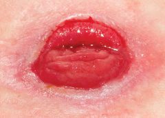
Venous ulcers are the most common type of lower extremity ulcer. They’re commonly found on the medial aspect of the lower extremity, from the ankle to the more proximal calf area. Usually, they arise along the course of the greater saphenous vein, but also may be lateral and may occur at multiple locations. They aren’t found above the knee or on the forefoot. Venous ulcers are shallower than arterial ulcers and have considerable exudate consistent with drainage from a ruptured water balloon. They may extend completely around the leg.
CVI: From a heavy sensation to visible changes
In patients with CVI, blood flows within a lower extremity in an abnormal, reverse direction, causing build-up of blood and intravascular fluid around the ankle. Initially, this may cause only a sensation of heavy legs toward the end of the day, with no visible changes. Eventually, it may lead to venous ulcers or other visible changes. This abnormal blood flow results from dysfunction of the normal mechanisms that drain blood from the leg against gravity into the IVC.
Sardina D. Skin and Wound Management Course; Seminar Workbook. Wound Care Education Institute; 2011:92-112.
Donald A. Wollheim is a practicing wound care physician in southeastern Wisconsin. He also is an instructor for Wound Care Education Institute and Madison College. He serves on the Editorial Board for Wound Care Advisor.
The number of people with diabetes who are meeting the ABC goals—hemoglobin A1C, blood pressure, and LDL cholesterol—has risen significantly in recent years, according to a study published by Diabetes Care. Patients meeting all three goals rose from about 2% in 1988 to about 19% in 2010.
Gains were made in each of the ABC goals, based on 2007 to 2010 data: 53% of patients met A1C goals, compared to 43% in 1988 to 1994 data; 51% met blood pressure goals, compared to 33%; and 56% met LDL goals, compared to 10%.
Younger people were less likely to meet A1C and cholesterol goals. Compared with non-
Hispanic whites, Mexican Americans were less likely to meet A1C and LDL goals and non-Hispanic blacks were less likely to meet blood pressure and LDL goals.
The researchers analyzed data from the National Health and Nutrition Examination Surveys from 1988–1994, 1999–2002, 2003–2006, and 2007–2010. Nearly 5,000 people age 20 or older participated.
Although progress had been made, the researchers conclude, “Despite significant improvement during the past decade, achieving the ABC goals remains suboptimal among adults with diabetes, particularly in some minority groups.”
Daily bathing with chlorhexidine-impregnated washcloths reduces infection risk
A study in The New England Journal of Medicine reports that daily bathing with chlorhexidine-impregnated washcloths reduces the risk of becoming infected with multidrug-resistant organisms and subsequent development of hospital-acquired bloodstream infections in intensive care unit patients.
“Effect of daily chlorhexidine bathing on hospital-acquired infection” included 7,727 patients in nine intensive care and bone marrow units in six hospitals. The units were randomly assigned to bathe patients with either no-rinse 2% chlorhexidine-impregnated washcloths or nonantimicrobial washcloths for 6 months; then, the units switched to the opposite product for 6 months.
The rate of infection with multidrug-resistant organisms was 23% lower in the chlorhexidine group and the rate of hospital-acquired bloodstream infection was 28% lower in the chlorhexidine group.
Patients tend not to wear custom-made footwear for preventing diabetic foot ulcers
Adherence to wearing prescription custom-made footwear was low among patients with diabetes, neuropathy, and a recently healed plantar foot ulcer, according to a study in Diabetes Care. The low adherence was particularly notable at home, where patients did the most walking.
Factors associated with higher adherence included lower body mass index, more severe foot deformity, and more appealing footwear.
Tedizolid works as well as linezolid in patients with acute bacterial skin infections
A JAMA study says that a 200-mg once-daily dose of oral tedizolid phosphate over 6 days was as effective as 600 mg of oral linezolid every 12 hours for 10 days in patients with acute bacterial skin and skin-structure infections, including cellulitis or erysipelas, major cutaneous abscesses, and wound infections.
A shorter course of tedizolid may be a “reasonable alternative” to linezolid for treating acute bacterial skin and skin-structure infections, the study concludes.
Water-based exercise improves ROM in patients with long-term arm lymphedema
A study of breast cancer survivors (median 10 years after surgery) with lymphedema found that a water-based exercise program improved shoulder range of motion (ROM).
The program consisted of at least twice-weekly water-based exercise for 8 weeks. At first, participants were supervised, but later they exercised independently. Although lymphedema status didn’t change, those who performed water-based exercise had an increase in ROM, showing improvement years after surgery.
Dehydrated amniotic membrane allograft possible option for treating chronic wounds
A dehydrated amniotic membrane allograft (EpiFix) was used to treat four patients whose wounds hadn’t closed after conservative and advanced measures and who had been referred for plastic procedures. A variety of wounds healed (located on the elbow, knee, hand, and ankle) after one to three applications of the amniotic material, which patients tolerated well. The wounds remained closed several months later.
Study casts doubt on MLD’s role in breast cancer–related lymphedema
A meta-analysis published in the World Journal of Surgical Oncology found the “current evidence” from randomized clinical trials “does not support” the use of manual lymphatic drainage (MLD) in preventing or treating lymphedema in patients with breast cancer.
The authors analyzed 10 randomized clinical trials with 566 patients.
CDC issues additional prevention steps for carbapenem-resistant Enterobacteriaceae
On Feb. 14, the Centers for Disease Control and Prevention (CDC) issued additional prevention steps for carbapenem-resistant Enterobacteriaceae (CRE). Increased reports of CRE prompted the action: Of the 37 unusual forms of CRE reported in the U.S., the last 15 have been reported since July 2012.
• When a CRE is identified in a patient with a history of an overnight stay in a healthcare facility (within the last 6 months) outside the U.S., send the isolate to a reference laboratory for confirmatory susceptibility testing and test to determine the carbapenem resistance mechanism.
• For patients admitted to healthcare facilities in the U.S. after recently being hospitalized (within the last 6 months) in countries outside the U.S., consider performing rectal screening cultures to detect CRE colonization, and place patients on contact precautions while awaiting the results.
Examples of Enterobacteriaceae include Klebsiella species and Escherichia coli. CRE are Enterobacteriaceae with high levels of resistance to antibiotics, including carbapenems. CRE infections most commonly occur among patients who are receiving antibiotics and significant medical treatment for other conditions.
At one time or another, all wound care professionals encounter a chronic wound, defined as a wound that fails to heal in an orderly and timely manner. Globally, about 67 million people (1% to 5% of the world’s population) suffer chronic wounds. In the United States, chronic wounds affect 6.5 million people and cost more than $25 billion annually to treat. (more…)
In the current healthcare environment, wound care practitioners need to capitalize on all available reimbursement avenues for care delivery and wound care supplies and dressings. And when it comes to reimbursement, there’s one constant: The rules change constantly. Whether these changes always benefit the patient is questionable. Nowhere is this more evident than in acute-care settings. Clinicians constantly are challenged to make sure their patient-care decisions comply with current Medicare reimbursement guidelines. (And if you’re not sure about today’s guidelines, be prepared for the guidelines to change tomorrow.) (more…)
Necrotizing fasciitis (NF) results from an infection that attacks the fascia and subcutaneous tissues. The primary bacterial etiology is group A streptococcus, a facultative anaerobic bacterium. However, other bacteria may contribute. Sometimes called the “flesh-eating” disease because of the potentially devastating effect on the afflicted patient, NF can be monomicrobial or polymicrobial.
The four typical settings for NF are:
surgical bowel or abdominal trauma surgery
pressure ulcer and perianal abscess
injection sites (especially in drug users)
Bartholin abscess or minor vulvovaginal infection.
Because of the rapid course and ravaging nature of acute NF, clinicians must maintain a high index of suspicion if the patient has suggestive signs and symptoms. In 1990, puppeteer Jim Henson (best known for creating the Muppets) died from NF. At that time, little was known about the progression of group A streptococcal infection.
The disease can quickly cause death, so starting immediate treatment is even more crucial than confirming the diagnosis. Once the disease is suspected, antibiotics must be given immediately and the patient must be prepared for surgery at once. NF spreads rapidly, capable of progressing from a small lesion to death in days to weeks. Thus, delayed diagnosis increases the risk of death. Lack of knowledge about the disease and inability to recognize it promptly are the main reasons many victims die. This article can improve your knowledge base.
Overview
NF was discovered in 1871 by Joseph Jones, a Confederate Army surgeon. At that time, it was called hemolytic streptococcal gangrene, nonclostridial gas gangrene, nonclostridial crepitant cellulitis, necrotizing or gangrenous erysipelas, necrotizing cellulitis, bacterial synergistic gangrene, or synergistic necrotizing cellulitis.
NF involves the fascia, muscle compartments, or both. It can affect not only the muscle fascia but the superficial fascia. NF and cellulitis differ in the amount of tissue involved and extent of tissue involvement.
The most common areas of infection are the abdominal wall, perineum, and extremities. When NF affects the perineum and scrotum, it’s called Fournier gangrene, after the French dermatologist and virologist Alfred Jean Fournier.
The most common causes are trauma, surgery, and insect bites. The disease can affect persons of any age. Such comorbidities as diabetes, chronic renal failure, immunosuppressive therapy, hypertension, obesity, and malnutrition increase susceptibility.
Pathophysiology
NF falls into four classifications based on wound microbiology. Type 1, the most common, involves polymicrobial bacteria. Type 2 results from trauma and is associated with comorbidities. Type 3, rare in this country, stems from gram-negative marine bacteria. Type 4 is a fungal infection occurring mostly in immunocompromised persons. (See Comparing types of necrotizing fasciitis by clicking the PDF icon above.)
Disease progression
The four types of NF progress in a similar way. Bacteria secrete pyrogenic exotoxin A, which stimulates cytokines. These cytokines damage the endothelial lining; fluid then leaks into the extravascular space.
M proteins in streptococci and β-hemolytic streptococci exacerbate the immune reaction by inhibiting phagocytosis of polymorphonuclear leukocytes and normal neutrophil chemotaxis. As the immune reaction increases, blood vessels dilate, allowing toxins to leak through vessel walls, which in turn decreases blood flow. As the cascade continues, hypoxic conditions cause facultative aerobic organisms to grow and become anaerobic. These bacteria exacerbate destruction of surrounding cells and lead to release of carbon dioxide, water, hydrogen, nitrogen, hydrogen sulfide, and methane. As the infection continues to progress, toxins spread throughout the bloodstream and the patient becomes septic.
Assessment
Obtain the patient’s medical history and description of the wound. Determine when the changes first appeared and whether the affected area seemed to get worse recently.
In all NF types, patients commonly present with a small, painful area (possibly with entry marks) but no other signs or symptoms. The wound may appear as a bulla, cellulitis, or dermatitis, representing an infection developing in underlying tissues. The skin may have a wooden-hard feel as the infection progresses to the subcutaneous space and causes necrosis. The wound becomes discolored and necrotic; drainage is rare until surgical debridement begins. The patient quickly develops fever, chills, nausea, and vomiting. As NF progresses, bullae become dark purple with darkened edges; the patient grows disoriented and lethargic, and organ failure and respiratory failure
ensue. Without treatment, the patient dies.
Diagnosis
Diagnostic tests usually include magnetic resonance imaging, complete blood count with differential, comprehensive metabolic panel, and cultures. (See Diagnostic findings in necrotizing fasciitis by clicking the PDF icon above.)
Treatment
Immediate surgical debridement and broad-spectrum antibiotics are needed to stop the immune response to infection. Clindamycin, gentamicin, penicillin, or metronidazole may be given alone or in combination until culture results are available. Supportive care includes total parenteral nutrition for nutritional support, I.V. fluids, and oxygen. Limb amputation should be done only as a last resort.
Surgical debridement involves penetrating deep into the fascia and removing all necrotic tissue. After the first debridement, release of “dishwater fluid” may occur.
Administering hyperbaric oxygen therapy (HBOT) after the first debridement increases tissue oxygenation, thus reducing tissue destruction by anaerobic bacteria. During HBOT (usually given as a 90-minute treatment), the patient breathes 100% oxygen in an environment of increasing atmospheric pressure.
HBOT should be given in conjunction with surgical debridement (usually after each debridement) and should continue until necrotic tissue ceases and cell destruction stops. HBOT also promotes collagen synthesis and neoangiogenesis (new blood vessel growth), which boosts blood supply and oxygen to tissues.
Adverse effects of HBOT include ear pain, oxygen toxicity, and seizures. Ear pain can be minimized by swallowing or yawning. If the patient continues to have ear pain, ear tubes may be inserted by an otolaryngologist. During HBOT, air breaks (intervals of breathing room air) are important in controlling oxygen toxicity (the main cause of seizures).
Throughout the HBOT treatment period, wound dressings must be simple. Well-moistened gauze dressings and an abdominal pad provide good support. Once necrotic destruction occurs, dressings depend on wound size and the need to fill cavities. The patient may require a diverting colostomy, depending on wound
location and the amount of uncontrolled diarrhea. Blood glucose levels must be monitored before and after HBOT, as this treatment affects blood glucose.
Supportive care and follow-up treatment
During initial treatment, patients need supportive care and monitoring. Once they’re out of danger, begin teaching them how to prevent NF recurrences. Advise them to control blood glucose levels, keeping the glycated hemoglobin (HbA1c) level to 7% or less. Caution patients to keep needles capped until use and not to reuse needles. Instruct them to clean the skin thoroughly before blood glucose testing or insulin injection, and to use alcohol pads to clean the area afterward.
Before discharge, help arrange the patient’s aftercare, including home health care for wound management and teaching, social services to promote adjustment to lifestyle changes and financial concerns, and physical therapy to help rebuild strength and promote the return to optimal physical health. One helpful patient resource is the National Necrotizing Fasciitis Foundation. The Centers for Disease Control and Prevention section on necrotizing fasciitis includes “Common sense and great wound care are the best ways to prevent a bacterial skin infection.”
The life-threatening nature of NF, scarring caused by the disease, and in some cases the need for limb amputation can alter the patient’s attitude and viewpoint, so be sure to take a holistic approach when dealing with the patient and family. Today, NF has a much better survival rate than 2 decades ago when Jim Henson died. In my practice, I’ve seen four NF cases. Thanks to early identification, good wound care, and HBOT, these patients suffered only minimal damage.
Selected references
Boyer A, Vargas F, Coste F, et al. Influence of surgical treatment timing on mortality from necrotizing soft tissue infections requiring intensive care management. Intensive Care Med. 2009;35(5):847-853. doi:10.1007/s00134-008-1373-4.
Cain S. Necrotizing fasciitis: recognition and care. Practice Nurs. 2010;21(6):297-302.
Centers for Disease Control and Prevention. Notes from the field: fatal fungal soft-tissue infections after a tornado—Joplin, Missouri, 2011. MMWR. 2011;60(29):992.
Chamber AC, Leaper DJ. Role of oxygen in wound healing: a review of evidence. J Wound Care. 2011; 20(4):160-164.
Christophoros K, Achilleas K, Vasilia D, et al. Postraumatic zygomycotic necrotizing abdominal wall fasciitis with intraabdominal invasion in a non immunosuppressed patient. Internet J Surg. 2007;11(1). doi:10.5580/17a8.
Ecker K-W, Baars A, Topfer J, Frank J. Necrotizing fasciitis of the perineum and the abdominal wall-surgical approach. Europ J Trauma Emerg Surg. 2008;
34(3):219-228. doi:10.1007/s00068-008-8072-2.
Hunter J, Quarterman C, Waseem M, Wills A. Diagnosis and management of necrotizing fasciitis. Br J Hosp Med. 2011;72(7):391-395.
Magel DC. The nurse’s role in managing necrotizing fasciitis. AORN J. 2008;88(6):977-982.
Phanzu MD, Bafende AE, Imposo BB, Meyers WM, Portaels F. Under treated necrotizing fasciitis masquerading as ulcerated edematous Mycobacterium ulcerans infection (Buruli ulcer). Am J Trop Med Hyg. 2012;82(3):478-481.
Ruth-Sahd LA, Gonzales M. Multiple dimensions of caring for a patient with acute necrotizing fasciitis. Dimens Crit Care Nurs. 2006;25(1):15-21.
Stevens DL, Bisno AL, Chambers HF, et al; Infectious Diseases Society of America. Practice guidelines for the diagnosis and management of skin and soft-tissue infections. Clin Infect Dis. 2005;41(10):1373-1406.
Su YC, Chen HW, Hong YC, Chen CT, et al. Laboratory risk indicator for necrotizing fasciitis score and the outcomes. ANZ J Surg. 2008;78(11):968-972.
Taviloglu K, Yanar H. Necrotizing fasciitis: strategies for diagnosis and management. World J Emerg Surg. 2007;2:19.
Lydia Meyers is a medical reviewer for National Government Services in Castleton, Indiana, and a clinical liaison at CTI Nutrition in Indianapolis. She has 11 years of wound care experience in nursing homes, wound clinics, and home health.