
The goal of wound-bed preparation is to create a stable, well-vascularized environment that aids healing of chronic wounds. Without proper preparation, even the most expensive wound-care products and devices are unlikely to produce positive outcomes.
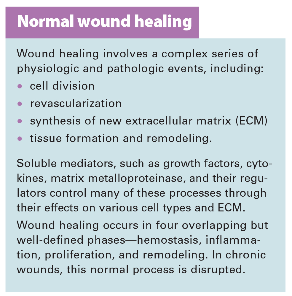
To best prepare the wound bed, you need to understand wound healing physiology and wound care basics, as well as how to evaluate the patient’s overall health and manage wounds that don’t respond to treatment. (See Normal wound healing.)
Basic wound care: DIME
To choose the right method of wound-bed preparation for a particular wound, first assess your patient’s condition, wound history, physical wound characteristics, and availability of resources. Local wound-bed preparation factors can be summarized by the acronym DIME—Debridement, Infection or Inflammation, Moisture balance, and Epithelial advancement. These four components address the various pathophysiologic abnormalities underlying chronic wounds.
D: Debridement
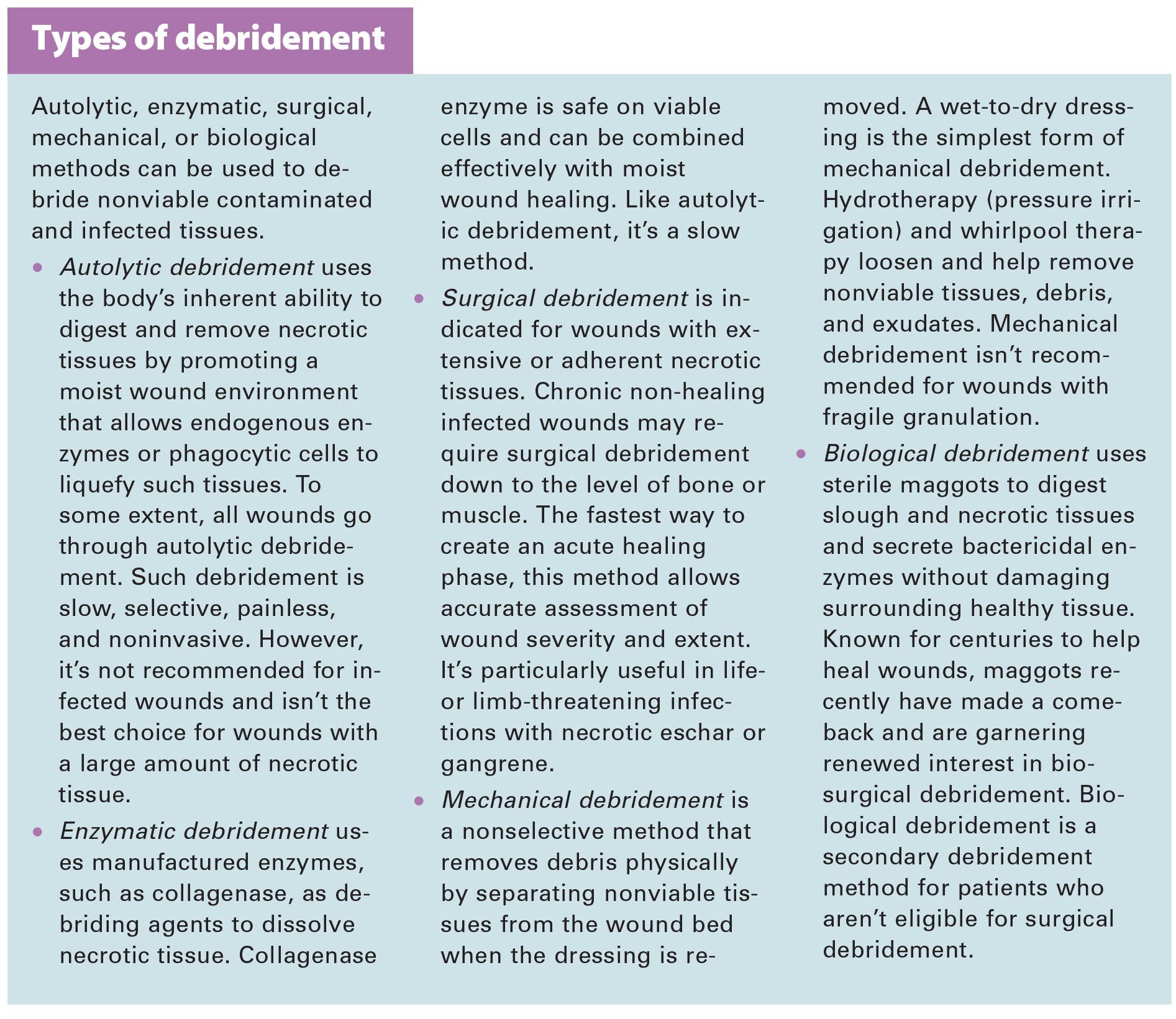
Nonviable tissue must be debrided because cell debris impairs healing. Research and clinical evidence show that debriding necrotic or fibrous tissue accelerates wound healing. (See Types of debridement.)
I: Infection or inflammation
Bacterial load directly impacts wound-bed preparation. Assess and treat the patient’s wound for superficial or serious infection, persistent inflammation, extensive colonization, and cellulitis. With infection or inflammation, wound healing stalls because the extracellular matrix and growth factors degrade more rapidly than they synthesize, impeding progression toward the proliferative phase and ultimately affecting re-epithelialization. Managing the bacterial load with local or systemic therapy is crucial to wound-bed preparation.
M: Moisture balance
Appropriate moisture promotes the action of growth factors and cytokines and aids migration of cells, including fibroblasts and keratinocytes. However, attaining a moisture balance is challenging. Excessive moisture can damage the wound bed and surrounding skin, leading to maceration and skin breakdown. Inadequate moisture, on the other hand, can impede cellular activities and promote eschar, resulting in poor wound healing.
Be sure to evaluate the patient’s nutritional status, cardiac and peripheral vascular status, and renal function. Check for risk factors that can cause moisture imbalance. Then identify an appropriate treatment plan. To increase compliance, explain planned interventions to patients and their caregivers. The typical plan includes medical wound management strategies, such as manual lymph drainage, compression devices and garments, absorptive dressings, and negative pressure wound therapy.
E: Epithelial advancement
Cellular dysfunction and biochemical imbalance can stall wound-bed progression by impeding epidermal cell and keratinocyte migration. Migration of epidermal cells and keratinocytes indicates the wound bed is adequately prepared. Wound contraction is another key sign of an adequately prepared wound bed.
Assessing the patient’s overall health
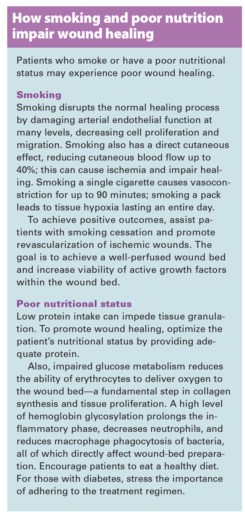
Remember—you must treat the patient before you can treat the wound. Especially with difficult wounds, multiple comorbidities can delay or interrupt re-epithelialization. Adequate nutrition and smoking cessation are especially important. (See How smoking and poor nutrition impair wound healing.)
Checking for wound-specific problems
Assess the patient for two wound-specific problems—biofilms and abnormal matrix.
• Biofilms delay wound healing by creating a host-pathogen environment that promotes cohabitation of many bacterial species and anaerobes. These bacteria promote their own survival within the wound environment. Elderly patients and those with complex diseases, diabetic foot ulcers, venous leg ulcers, or pressure ulcers may develop extensive bacterial populations. That’s why restoring bacterial balance is important in managing chronic wounds. Techniques for managing biofilms effectively include use of topical agents or systemic antibiotics and regular maintenance debridement.
• Abnormal matrix can develop in chronic wounds containing proteases that digest fibronectin and growth factors in the fibrin clot, causing a degraded matrix that no longer supports re-epithelialization or formation of granulation tissue.
Managing wounds unresponsive to treatment
Cells in chronic wounds become unresponsive, unable to divide or respond to such messengers as cytokines and growth factors. This results in phenotypic dysregulation. For successful wound-bed preparation, options may include bioengineered skin-cell therapy, stem-cell therapies, and platelet-rich plasma (PRP) for cutaneous wounds.
Bioengineered skin-cell therapy
Bioengineering treatments have provided viable therapeutic options, especially in managing chronic or difficult-to-heal wounds. Bioengineered skin and soft-tissue substitutes can be acellular or cellular.
Acellular products contain a matrix or scaffold composed of such materials as collagen, hyaluronic acid, and fibronectin. These products differ in various ways, including:
• species source (human, bovine, or porcine)
• tissue source (such as dermis, pericardium, or intestinal mucosa)
• additives (for instance, antibiotics or surfactants)
• hydration (wet or freeze-dried)
• required preparation (multiple rinses or rehydration).
Cellular products contain living cells, such as fibroblasts and keratinocytes within the matrix. Cells within the matrix may be autologous, allogeneic, or derived from other species (such as sheep or pigs). Skin substitutes also may be composed of dermal cells, epidermal cells, or a combination and may provide growth factors to stimulate healing. Topical growth factors are used as an adjuvant treatment to such therapies as debridement, off-loading, frequent dressing changes, and compression of wounds caused by vascular insufficiency
Stem-cell therapy
Use of stem cells in tissue regeneration has significant potential in cutaneous wound-bed preparation. Stem cells act in several ways to aid wound repair; in chronic wounds, they could serve as an additional tool for therapeutic augmentation. This combined mode of repair and regeneration probably explains why cell therapies are more effective than simpler alternatives, such as direct growth-factor therapy treatment.
PRP
Multiple studies since the 1990s have shown promising results for PRP. For instance, researchers found PRP gel accelerates bone regeneration and studies confirmed the presence of specific platelet-derived growth factor receptors (PDGFs) in bone tissue. Research has uncovered strong evidence that these therapies are effective.
The PRP gel aids molecular and cellular induction of normal wound-healing responses, similar to platelet activation. In addition, PRP contains several growth factors and other cytokines that stimulate healing of bone and soft tissues. Autologous PRP is an advanced wound therapy used for hard-to-heal acute and chronic wounds.
Growth factors such as topical PDGF can help correct matrix abnormalities of growth factors. Also, studies have found that accelerated wound healing follows topical application of epidermal growth factor derived from the patient’s own cultured keratino cytes, delivered in a fluid compress.
A systematic approach
When preparing the patient’s wound bed and managing the wound, use a systematic approach that helps remove barriers to healing. Proper preparation helps ensure optimal patient outcomes.
Kulbir Dhillon is a nurse practitioner and wound care specialist with VA HBPC in Sacramento, California.
Selected references
Falanga V. Wound bed preparation: science applied to practice. In: Wound Bed Preparation in Practice. EWMA Position Document. London: Medical Education Partnership Ltd;2004;2-5.
Fitzgerald RH, Armstrong DG. Palliative advanced wound care. Vein Mag. 2009;2(2):44-6.
Harris RJ. The nursing practice of conservative sharp wound debridement: promotion, education and proficiency. Wound Care Canada. 2009;7(1):22-30.
Hess CT. Checklist for factors affecting wound healing. Adv Skin Wound Care. 2011;24(4):192.
Knox KR, Datiashvili RO, Granick MS. Surgical wound bed preparation of chronic and acute wounds. Clin Plast Surg. 2007;34(4):633-41.
Laplaud AL, Blaizot X, Gaillard C, et al. Wound debridement: comparative reliability of three methods for measuring fibrin percentage in chronic wounds. Wound Repair Regen. 2010;18(1):13-20.
Ligresti C, Bo F. Wound bed preparation of difficult wounds: an evolution of the principles of TIME. Int Wound J. 2007;4(1):21-9.
Mustoe TA, O’Shaughnessy K, Kloeters O. Chronic wound pathogenesis and current treatment strategies: a unifying hypothesis. Plast Reconstr Surg. 2006;117(7 Suppl):35S-41S.
Opletalová K, Blaizot X, Mourgeon B, et al. Maggot therapy for wound debridement: a randomized multicenter trial. Arch Dermatol. 2012;148(4):432-8.
Panuncialman J, Falanga V. The science of wound bed preparation. Surg Clin North Am. 2009;89(3):611-26.
Paul AG, Ahmad NW, Lee HL, et al. Maggot debridement therapy with Lucilia cuprina: a comparison with conventional debridement in diabetic foot ulcers. Int Wound J. 2009;6(1):39-46.
Payne WG, Salas RE, Ko F, et al. Enzymatic debriding agents are safe in wounds with high bacterial bioburdens and stimulate healing. Eplasty. 2008;7;8:e17.
Sardina D. Wound bed preparation. In: Skin and Wound Management Course Workbook. Lake Geneva, WI: Wound Care Education Institute; August 2010;5:42-58.
Sibbald RG, Goodman L, Woo KY, et al. Special considerations in wound bed preparation 2011: an update. Adv Skin Wound Care. 2011;24(9):415-36.
Sibbald RG, Orsted HL, Schultz GS, et al. Preparing the wound bed 2003: focus on infection and inflammation. Ostomy Wound Manage. 2003;49(11):23-51.
Wolcott RD, Rumbaugh KP, James G. Biofilm maturity studies indicate sharp debridement opens a time-dependent therapeutic window. J Wound Care. 2010;19(8):320-8.