As a wound care specialist, you have learned about many skin conditions, some so unusual and rare that you probably thought you would never observe them. I’ve been a nurse for 38 years, with the last 10 years in wound care, and that’s certainly what I thought. But I was wrong. Let me tell you about my challenging patient with an unusual skin condition.
A perplexing patient
Mrs. Thompson*, a 77-year-old resident in a long-term care facility where I work, had diabetes, peripheral vascular disease, and a history of a cerebrovascular accident in 1993, which left her with left-sided paralysis.
In February 2010, Mrs. Thompson underwent a colostomy in her left lower abdominal quadrant as a result of a large sigmoid colon volvulus. She was doing well until November 2011, when periostomal skin breakdown began, presumably caused by leakage. Over the course of the next 18 months, her skin breakdown would often improve without any change in treatment, which made subsequent exacerbations frustrating.
Here are the appliance-related adaptations my colleagues and I tried with Mrs. Thompson:
• stoma powders and paste
• wafer adaptations
• plain and medical-grade honey hydrocolloid applied directly to peristomal lesions.
Unfortunately, none of these efforts solved the problem. A March 2013 dermatology consult resulted in no definitive diagnosis or alternative treatment options.
My “Aha” moment came when a computer search for causes of periostomal breakdown revealed illustrations of various conditions. One image, labeled pyoderma gangrenosum (PG), resembled what was occurring with Mrs. Thompson.
About pyoderma gangrenosum
PG, a skin ulceration, was first described in 1930 by Brunsting and colleagues. It’s associated with Crohn’s or inflammatory bowel disease, cancer, blood dyscrasias, diabetes, and hepatitis.
PG has been described in several forms, but ulceration usually occurs on the abdomen, perineum, and lower extremities. The lesions begin as discrete pustules that erupt and coalesce into a classic painful ulcer with a violaceous border and undermined edge. Multiple lesions are common.
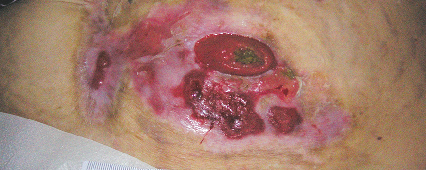
Mrs. Thompson’s wound at the time of diagnosis
Diagnosis
The diagnosis of peristomal PG is based on the patient’s history and characteristics of skin breakdown because biopsies and cultures can’t confirm the diagnosis. The lesions are typically very painful, but Mrs. Thompson didn’t experience pain because of the left-sided sensory deficits caused by her stroke. In one study, the time to onset of periostomal PG after creation of a stoma ranged from 2 months to 25 years; in Mrs. Thompson’s case, onset was 20 months. The erratic progression of this rare disease is considered a hallmark of the disorder.
My colleagues and I validated the diagnosis of periostomal PG by characteristics of the lesions and exclusion of other skin conditions.
Management
The unknown etiology of the peristomal lesions makes treatment decisions challenging. Because the condition is rare, recent evidence-based practice data are limited, with most reported as part of research trials. When lesions are mild and there is absence of systemic disease, it may be possible to control the condition with topical corticosteroids and dressings.
Based on what we could find in the literature and discussion with the geriatric nurse practitioner and Mrs. Thompson’s primary care physician, we decided to start her on high-dose steroid cream.
Positive results
On April 4, 2013, Mrs. Thompson began receiving daily clobetasol propionate 0.05%, a high-dose steroid cream applied to the peristomal area. We gently rubbed in the cream completely, followed by an aerosol skin barrier and a one-piece appliance. The treatment was re-evaluated every 14 days, as recommended by the manufacturer, because of the risk for hyperglycemia, which did not occur.
By June 13, 2013, 72 days later, the lesions had healed and we resumed biweekly appliance changes.
Lesions healed after 72 days of clobetasol
propionate 0.05%.
This healing time of about 2.4 months was much faster than what we found in the literature: One study reported an average healing time for periostomal PG of 11.4 months (median, 7.5 months; range 1-41 months).
After the initial PG exacerbation was healed, Mrs. Thompson occasionally experienced minor exacerbations, but not to the extent it was when first diagnosed and treated with clobetasol.
One 60-g tube of clobetasol propionate 0.05% (cost of $328) was required to successfully treat the breakdown.
Complete wound healing at 120 days
Committed to healing
Comorbid conditions play an important role in effectively diagnosing and treating skin breakdown. In Mrs. Thompson’s case, sensory deficits from a stroke sustained almost 20 years earlier diminished her ability to feel pain at the stoma site, which is the signature diagnostic characteristic of PG.
I learned that even though diagnosis of skin conditions can be difficult, it’s important not to give up. My commitment, along with the commitment of my colleagues, resulted in our ability to find a solution for Mrs. Thompson’s condition.
*Name is fictitious.
Susan Lee is a wound care provider for two long-term care facilities, Maluhia and Leahi Hospital in the Oahu Region of the Hawaii Health System Corporation in Honolulu, Hawaii.
Selected references
Alvey B, Beck DE. Peristomal dermatology. Clin Colon Rectal Surg. 2008;21(1):41-4.
Bhat RM. Pyoderma gangrenosum: an update. Indian Dematol Online J. 2012;3(1):7-13.
Brusting LA, Goeckerman WH, O’Leary PA. Pyoderma gangrenosum: clinical and experimental observations in 5 cases occurring in adults. Arch Dermatol. 1930;22:655-80.
Butcher M. Pydoerma gangrenosum: a diagnosis
not to be missed. Wound Care UK. 2005;1(3). http://www.wounds-uk.com/pdf/content_9031.pdf
Crowson AN, Mihm MC Jr, Magro C. Pyoderma gangrenosum: a review. J Cutan Pathol. 2003;30(2):
97–107.
Gray M, Catanzaro J. What interventions are effective for managing peristomal pyoderma gangrenosum? J Wound Ostomy Continence Nurs. 2004;
31(5)249-55.
Hughes AP, Jackson JM, Callen JP. Clinical features and treatment of peristomal pyoderma gangrenosum. JAMA. 2000;284(12):1546-8.
Sheldon DG, Sawchuk LL, Kozarek R, et al. Twenty cases of peristomal pyoderma gangrenosum. Arch Surg. 2000;135(5),564-9.
Wollina U. Pyoderma gangrenosum—a review. Orphanet J Rare Dis. 2007;2:19.